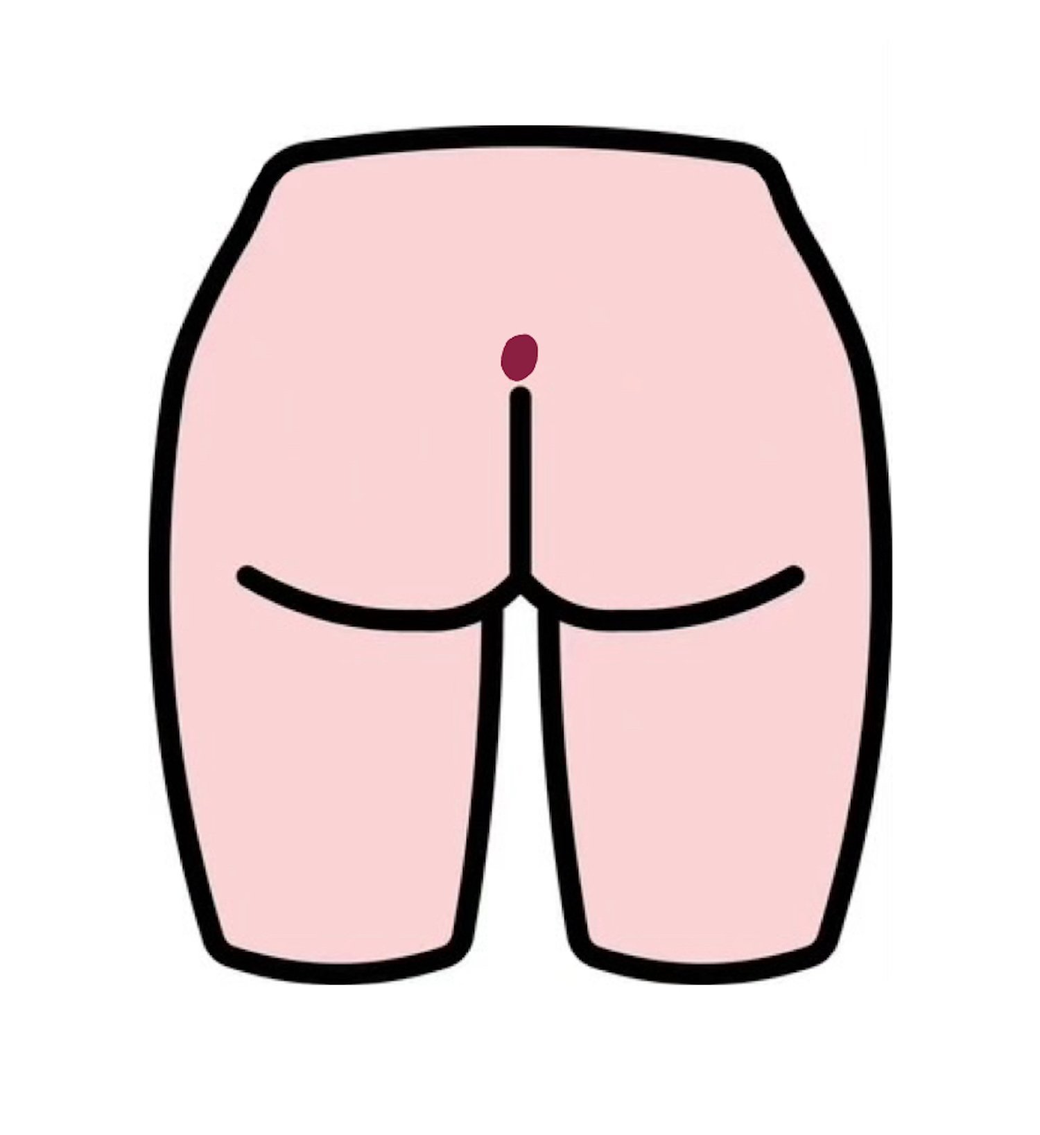
Pilonidal disease encompasses a spectrum of disease that ranges from simple asymptomatic sinuses to large open complex wound. The term derives its name from Latin pilus (hair) and nidus (nest). O.H. Mayo was the first to describe the disease in 1883.
Etiology
- Risk factors: male sex, family history, overweight/obese, trauma, irritation, sedentary lifestyle, hirsute habitus, poor hygiene
Pathogenesis
- Heavily debated
- Shed hair is drawn into the gluteal cleft by the motion from the buttocks, which creates a vacuum effect forcing the hair into the skin. The foreign body reaction produced by trapped hair may lead to a hair-filled abscess cavity, which can drain spontaneously through the skin or back through the sinus tracts.
Presentation
- Acute abscess cephalad to the gluteal cleft
- ± Pain and drainage
- Sinus openings along the midline of the gluteal cleft, 4-8 cm from the anus → hallmark finding
Diagnosis
- Clinical
- Dye (e.g., methylene blue) may be injected for better identification of complicated sinuses. It must be done several days before surgery to avoid excessive staining of the operative area.
Treatment
- Nonoperative management
- Feasible option for minor disease
- Shaving
- Hygiene practices
- Weight loss
- Avoid prolonged sitting
- Surgery
- Simple procedures
- Abscess drainage
- Indicated for acute pilonidal abscess with active infection
- Incise abscess cavity just off of midline and break up all loculations to drain purulence entirely
- Pack wound initially and remove the following day as there is no evidence to support continued packing
- Shower 1-2 times daily to keep area cleanWound usually closes in 1-2 weeks
- Non-excisional procedures
- Indicated for minor disease
- Remove hair and debris from pits
- Inject pits with phenol (or fibrin glue) to create epithelial destruction and inflammation to aid in closure of the tract
- Pit-picking
- Indicated for mild chronic disease with main complaint of pits
- Central pits are excised using punch knife slightly larger than the pit. Knife is inserted and full depth of pit including hair and debris are removed and wound is closed primarily with 3-0 Vicryl
- Pits usually close in 1-3 weeks
- Excision ± primary wound closure
- Gold standard
- Rarely results in complex wound
- Abscess drainage
- Intermediate procedures → mild to moderate disease
- Karydakis flap
- Excision of affected midline tissue
- Creation of skin flap with beveled edges to cross midline
- Tension-appropriate primary closure in layers to obliterate dead space
- Cleft lip procedure
- Originally coined by Dr. John Bascom
- Tissue marked where gluteal tissue joins at midline followed by taping the buttocks apart to reveal a “wishbone” shaped mark which establishes a “safe zone” beyond which no further dissection should be completed.
- Additional elliptical mark is created over area with more significant disease after skin is prepped.
- Skin flap is created by dissecting toward the opposite side safe-zone boundary and mobilized across the midline. Wound is closed in layers to obliterate dead space.
- Karydakis flap
- Complex procedures → complex/destructive disease, almost always require closed-suction drainage
- Rhomboid flap/Limberg flap
- Excise all disease with a diamond-shaped to level of post-sacral fascia
- Helpful to orient rhombus shaped slightly counterclockwise
- Flap is mobilized from donor site (usually right buttock)
- Wound is closed in layers
- No strenuous physical activity for 4-6 weeks
- Rhomboid flap/Limberg flap
- Simple procedures
Relevant Information
- The location of a pilonidal cyst is different from a perirectal abscess. Perirectal abscesses are found near the anus.
- Principles of intervention
- Use off-midline excision and closure to aid with healing
- Tension-appropriate repair
- Flatten natal cleft
Complications
- Recurrence
- Wound breakdown
- Infection
Differential Diagnoses
- Abscess
- Hidradenitis suppurativa
- Inflammatory bowel disease
- Anal fistula
- Epidural abscess
- Folliculitis
- Furuncles
- Carbuncles