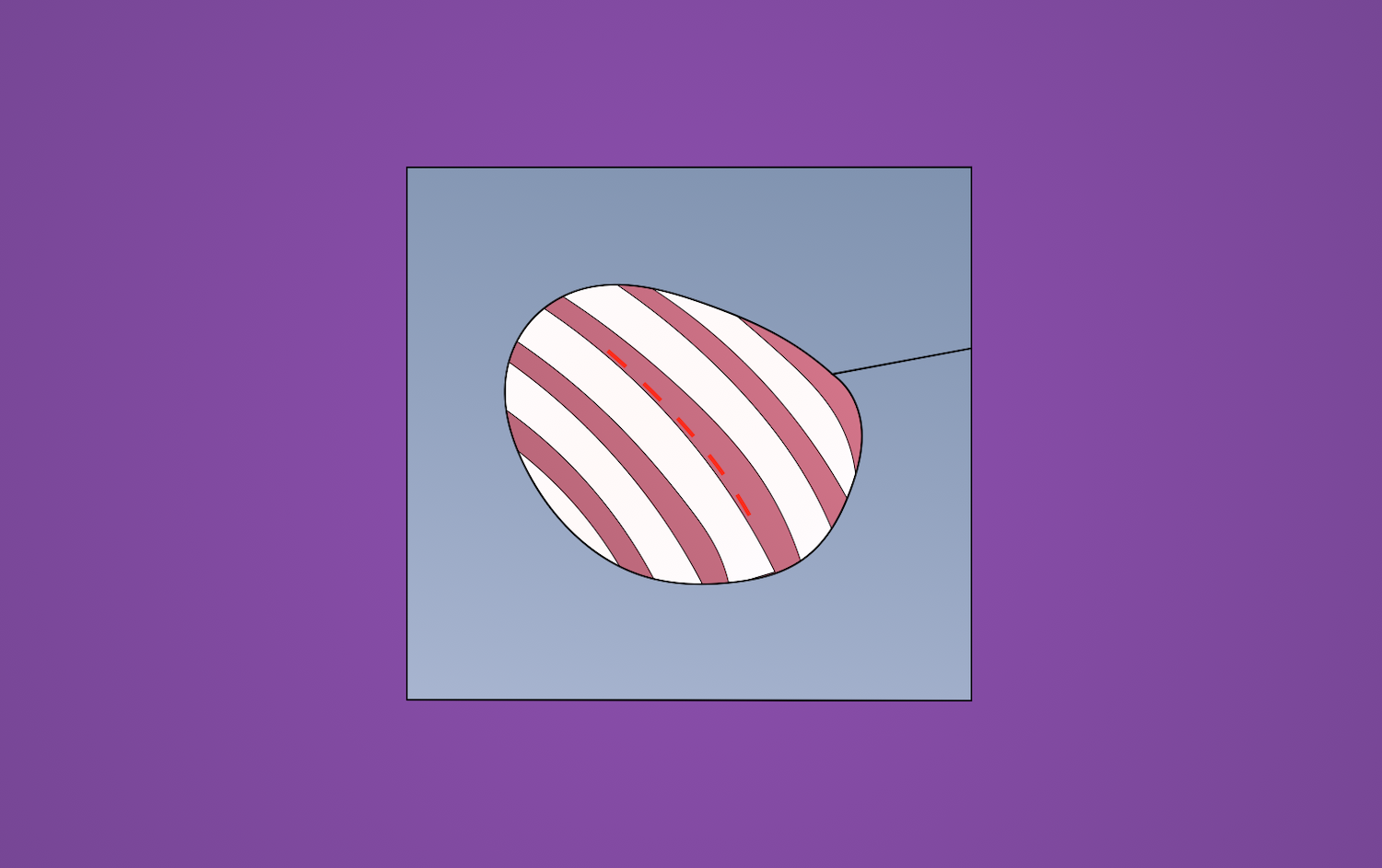
Thoracotomy is a surgical procedure in which an incision is made between the ribs to gain access to organs in the chest/thorax. Emergent thoracotomy first came to the United States in the late 1800s/early 1900s. In 1873, Moritz Schiff, a German physiologist, proposed the use of thoracotomy for open cardiac massage.
Indications
- Lung biopsy or lung cancer
- Cardiovascular conditions
- Diaphragm conditions
- Pneumothorax
- Cardiac tamponade
- Esophageal diseases or esophageal cancer
- Pleural effusion
- Emergent procedures
Preoperative Considerations
- Preventative spirometry, to improve compliance postoperatively
- Smoking cessation *** weeks prior to elective procedure
- Pulmonary function testing
- Room air arterial arterial blood gas
- Preoperative pulmonary rehabilitation, as indicated
Relevant Anatomy
- Auscultatory triangle: superior border of latissimus dorsi, inferior border of the trapezius, and medial border of scapula
- Trapezius
- Action: scapula rotation, retraction, elevation, depression
- Nerve: spinal accessory (CN XI)
- Latissimus dorsi
- Action: adduction, extension, internal rotation of the arm
- Nerve: thoracodorsal nerve (C6-C8)
- Rhomboid
- Action: rotate scapula, medial movement
- Nerve: dorsal scapular
- Serratus anterior
- Action: scapula protraction, stabilization, upward rotation
- Nerve: long thoracic (C5-C7)
Relevant Information
- A patient able to walk at least three flights of stairs will tolerate thoracotomy
Surgical Technique
- Prior to thoracotomy, fibrooptic bronchoscopy is performed through single-lumen ET tube to remove secretions, visualize anatomy, and screen for masses
- Patient is placed in the lateral decubitus position with hips secured to the operating table with tape. The lower leg is flexed at the knee and a pillow is placed between it and the extended upper leg. An axillary roll is placed to support the shoulder and upper thorax on the side contralateral to the thoracotomy, with the arm extended forward perpendicular to the operating table. The arm on the side of the thoracotomy is extended forward and upward and placed on an arm holder in line with the head. Skin is cleaned and prepped in the usual sterile fashion.
- The incision is made with the surgeon standing posterior to the patient. Various incisions can be made to accomplish a thoracotomy, including a posterior, axillary, or anterior incision.
- Posterior: oblique incision with or without sparing latissimus dorsi with the chest entered through fourth or fifth interspace; for pulmonary resection
- Vertical axillary: incision made anterior to latissimus dorsi with chest entered through fourth interspace; for hilar visualization
- Anterior or anterolateral: curvilinear incision under inferior border of pectoralis major at inframammary fold
- Medial sternotomy: vertical incision from sternal notch to xiphoid with bone saw to divide sternum at midline; access to mediastinum, heart, great vessels, and right and left hemithorax
- Transverse/clamshell: longer incision than median sternotomy – combines two anterior thoracotomy incisions in inframammary fold with transverse division of sternum at fourth intercostal space with ligation of both internal mammary arteries; access left and right hilum
- Verification that the patient is on single lumen ventilation is accomplished, and an incision is made entering the pleura. The lung then drops away allowing the incision to be extended to the desired length. Alternatively, the rib is transected at the costal cartilage of the neck with rib shears and a self-retaining retractor is placed and opened gradually.
- Chest tube(s). Chest tubes are usually placed (and must be at least 32 French in size). Two chest tubes (or one, if desired) may be placed, with one over the diaphragm in the posterior gutter along the spine and the other directed anteriorly. The posterolateral chest tube is brought out through stab wounds in the skin as low as possible, and ideally anterior to the midaxillary line.
- Closing. Encircling 1-0 absorbable sutures are placed and can be tied with or without a rib approximator. If ribs were transected or fractured during spreading, sutures must encircle both ribs and immobilize all rib fragments. Placing a suture through the sacrospinalis muscle and fixing it to the neck of the transected rib and rib above can help achieve hemostasis. Chest muscles are approximated using running or interrupted absorbable suture, with care to approximate the layers separately (rhomboids, serratus anterior, trapezius, latissimus dorsi). Subcutaneous 3-0 nonabsorbable sutures prevent incision disruption when skin staples are removed on postoperative day 7-8.
Postoperative Considerations
- Incentive spirometry
Postoperative Complications
- Bleeding
- Infection
- Pneumothorax
- Pleural effusion
- Shoulder dysfunction
- Pain
- Post-thoracotomy pain syndrome
