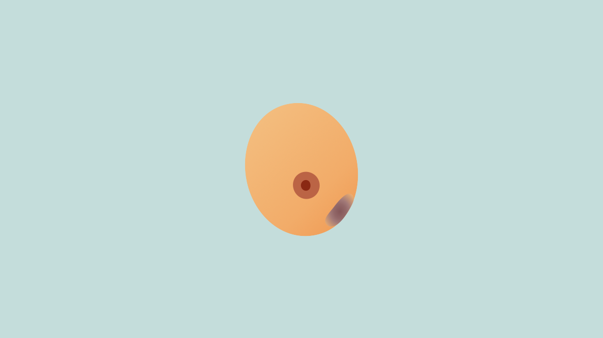
Breast fat necrosis was described by Geoffrey Hadfield in a report written in 1926 in the British Journal of Surgery. In this description, it was described as an “innocent condition” that could mimic breast cancer. Prior to this, Lee and Adair had described the condition in 1920.
Etiology
- Idiopathic (most common)
- Surgery for benign/malignant diseases
- Breast reconstruction
- Lipomodelling
- Radiation
Pathogenesis
- Fat cells damaged by iatrogenic trauma
- After this, the body creates scar tissue to repair the damaged tissue
- In some cases, fat cells die and release their contents forming fluid-filled collections that often have circumferential “eggshell” calcification called “oil cysts”
Presentation
- Usually clinically occult, however can be a palpable mass when clinically evident
- Firm round lump
- Usually painless
- Skin dimpling
- Nipple retraction
- May have overlying ecchymosis, skin thickening, erythema, or retraction
- Often seen in superficial areas of breast (due to adipocytes and surrounding microvessels)
Imaging
- Breast ultrasound → round/oval, smooth-bordered, hypoechoic or anechoic, may see calcifications which appear more hyperechoic associated with acoustic shadowing
- Mammogram → round/oval, smooth bordered, lucent mass with thin rim
- Histology → lipid-laden macrophages, scar tissue, and chronic inflammatory cells
Treatment
- Nodule at area of fat grafting, subcutaneous, <0.5 cm without skin changes → monitor with serial exams; perform ultrasound if it changes
- Nodule outside area of fat grafting → ultrasound with biopsy
- Firm nodule → ultrasound with biopsy
- Nodule >0.5 cm with associated skin changes → ultrasound with biopsy
- Ultrasound confirming the area in concern is fat necrosis → observation with repeat ultrasound in 6 months
Relevant Information
- No malignant potential
