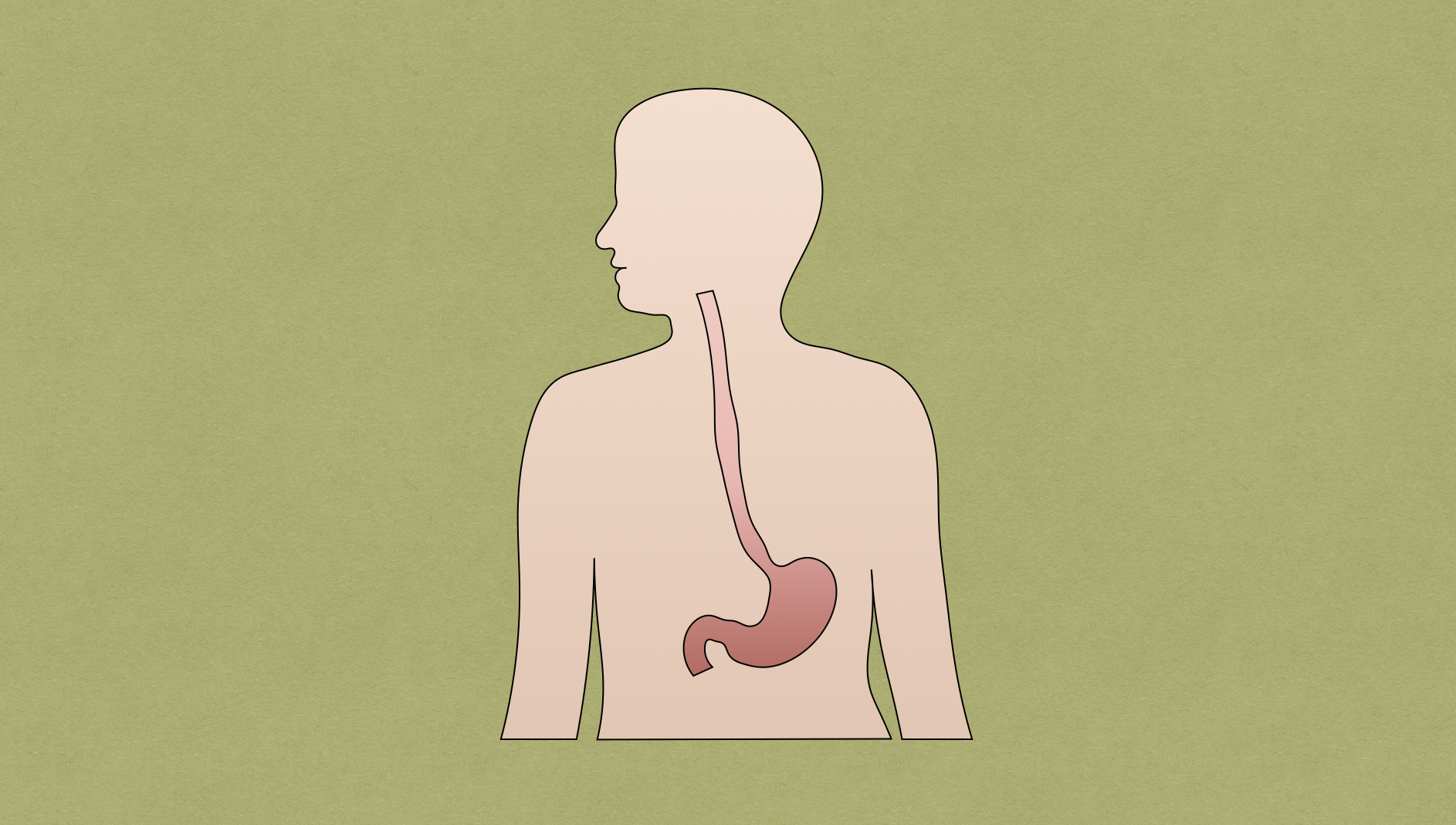
Andreas Vesalius was an anatomist and physician who was the first person to provide an anatomical description of the esophagus in 1543 in his book titled De Humani Corporis Fabrica. Thomas Billroth was the first person to attempt surgery on the neck, which was attempted in 1871 in dogs. This was later carried out in people by Vincenz Czerny in 1877.
Pathway and Segments
- Two-layered, mucosa-lined muscular tube measuring roughly 25-30 cm in length
- Begins in the neck at the pharynx (at C6) and ends in the abdomen where it joins with cardia of stomach (at T11)
- Takes a winding course, beginning as midline structure and deviates slightly to the left at the trachea as it passes through the neck into the thoracic inlet. Then at the carina it deviates to the right to accommodate the aortic arch prior to winding back to the left as it goes under left mainstem bronchus and is pushed anteriorly by the descending thoracic aorta before entering the diaphragm.
- Esophageal segments
- Pharyngeal
- Cervical
- Thoracic
- Abdominal
Esophageal Layers
- Mucosa (innermost layer) → made of squamous epithelium
- Epithelium
- Basement membrane
- Lamina propria
- Muscularis mucosae
- Submucosa
- Muscularis propria
- Upper ⅓ → striated
- Lower ⅔ → smooth
- Lacks serosa → cancer spreads through lymphatics
Sphincters
- Upper esophageal sphincter (UES)
- High-pressure zone at inlet of esophagus → corresponds reliably to cricopharyngeus muscle
- Killian triangle
- Transition between oblique fibers of thyropharyngeus muscle and horizontal fibers of cricopharyngeus muscle
- Cricopharyngeus: generates high-pressure; marks position of UES and esophageal introitus
- Point of potential weakness → site of Zenker diverticulum
- Transition between oblique fibers of thyropharyngeus muscle and horizontal fibers of cricopharyngeus muscle
- Most common site of iatrogenic perforation and foreign body
- Innervation: recurrent laryngeal
- Lower esophageal sphincter (LES)
- Not true anatomic sphincter → high resting tone, relaxes with swallowing or gastric distention
- Prevents reflux
- Innervation: vagus
Anatomic Narrowing
- Cricopharyngeus muscle (narrowest point)
- Just below carina, where left mainstem bronchus and aorta abut esophagus
- Diaphragmatic constriction
Vasculature
- Cervical esophagus
- Supply: inferior thyroid arteries (branch of thyrocervical trunk on left and subclavian on right)
- Drainage: submucosal venous plexus → inferior thyroid vein → left subclavian and right brachiocephalic vein
- Cricopharyngeus muscle
- Supply: superior thyroid artery
- Drainage:
- Thoracic esophagus
- Supply: 4-6 esophageal arteries coming off of aorta and right and left bronchial arteries
- Drainage: submucosal venous plexus joint with esophageal venous plexus → azygos and hemiazygos veins
- Abdominal esophagus
- Supply: left gastric artery and paired inferior phrenic arteries
- Drainage: paired phrenic veins and left gastric veins and short gastric veins → systemic and portal venous systems
Lymphatics
- Arise from submucosa and muscularis layers
- Upper ⅔: superior
- Cervical esophagus: deep cervical lymph nodes
- Thoracic esophagus: paraesophageal and mediastinal lymph nodes
- Lower ⅓: inferior
- Abdominal esophagus: gastric/cardiac and celiac lymph nodes
- Drain into cisterna chyli/thoracic duct
Nerves
- Right vagus
- Twists over posterior aspect
- Innervates celiac plexus
- Left vagus
- Twists over anterior aspect
- Innervates liver and biliary tree
