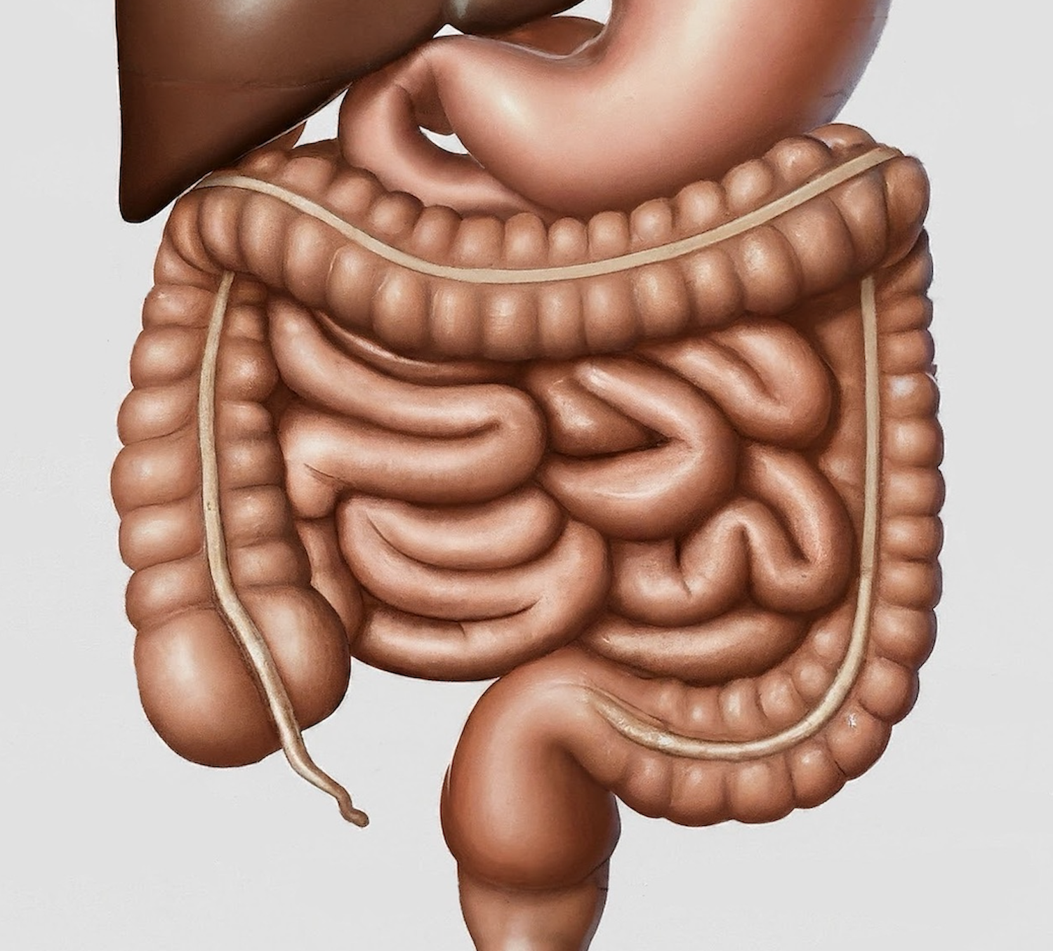
Inflammatory bowel disease is a chronic inflammatory disease of the gastrointestinal tract. It can be further divided into ulcerative colitis (UC) and Crohn disease.
The name “ulcerative colitis” was coined by Samuel Wils in 1859. Prior to this, a report by Matthew Baillie in 1793 described a case in which a patient died from what is now known as ulcerative colitis. Ileitis terminalis was first described in 1904 by Antoni Leśniowski. Burrill Bernard Crohn later submitted fourteen cases in 1932. Later in the same year, Crohn, along with Leon Ginzburg and Gordon Oppenheimer, published a case series on regional ileitis. The disease became known as Crohn disease due to the alphabetical order of the authors.
Etiology
- Protective factors: smoking (UC)
- Risk factors: genetics, environmental factors, smoking (Crohn)
Pathogenesis

History
- Diarrhea, usually bloody in UC
- Abdominal pain
- Tenesmus
- Urgency
- Extraintestinal manifestations (slightly higher prevalence in Crohn)
- Arthritis (most common), sacroiliitis
- Ankylosing spondylitis (HLA-B27 positive)
- Erythema nodosum: red, painful swollen nodules
- Pyoderma gangrenosum: extremely painful ulcerating lesions, usually occur at sites of repeated trauma
- Uveitis
- Iritis
- Episcleritis
- Sclerosing cholangitis
- UC may present with LLQ pain
- Crohn may present with RLQ pain
Physical exam
- Tachycardia
- Fever
- Pallor, depending on anemia
- Severe pain, fever, abdominal distention, chills, lethargy → suspicious for toxic megacolon
- Occult blood on digital rectal exam
- Crohn
- Diarrhea
- Weight loss
- Palpable tender abdominal mass
- External fistula
- Perianal disease (25%): anal fissure, anal skin tags, anorectal abscesses, anal stenosis
Labs
- Stool cultures (rule out infectious causes)
- Leukocytosis
- Thrombocytosis
- Elevated ESR, CRP
- Fecal calprotectin
Imaging
- Abdominal X-ray: assess for free air, bowel obstruction, or toxic megacolon
- Barium studies
- US, CT, MRI have been used in the diagnosis of inflammatory bowel disease or to assess for complications
- Endoscopy
- Essential to obtaining biopsies and confirming inflammatory bowel disease diagnosis
- Can be rigid proctoscopy, flexible sigmoidoscopy, or colonoscopy
- UC
- Inflammation beginning at the dentate line extending proximally
- Mayo Clinic Scoring System can be used for endoscopic assessment of UC
- Grade 1: normal endoscopic appearance
- Grade 2: slightly more erythematous
- Grade 3: even more erythematous area with touch bleeding
- Grade 4: significant bleeding and friability
- Crohn
- Inflammation is patchy, with discontinuous inflammation (i.e., skip areas) with areas of intervening normal-appearing mucosa
- Deeper punched-out ulcerations
- Most common site of involvement is ileocolic
Treatment
- Medical management
- Aminosalicylates: sulfasalazine, mesalamine
- Corticosteroids: used if patient has severe disease; prednisone, hydrocortisone
- Immunomodulators: thiopurines (e.g., azathioprine, 6-mercaptopurine), methotrexate
- Biologics: monoclonal antibodies against TNF-ɑ (e.g., infliximab)
- Surgery
- Indications
- Indications for surgery in UC
- Failure to respond to maximum medical therapy
- Severe disease, with multiple bowel movements, poor nutritional status, “failure to thrive,” need for surgery to regain good physical health; poor quality of life with urgency, tenesmus, low body weight
- Fulminant colitis; need hospitalized and placed on IV steroids
- Significant GI bleeding
- Children with failure to grow
- Presence of dysplasia or cancer
- Failure to respond to maximum medical therapy
- Indications for surgery in Crohn
- Symptoms of obstruction secondary to fibrostenosing Crohn disease
- Perforating Crohn disease with abscess or fistula
- Presence of many types of fistulas (relative indication)
- Significant abdominal pain associated with obstruction
- Children with failure to grow
- Presence of dysplasia or cancer
- Indications for surgery in UC
- Surgical options
- Surgical options for UC
- Subtotal colectomy and ileorectal anastomosis
- Ileostomy
- Hartmann procedure
- Total proctocolectomy with end ileostomy
- Proctocolectomy with either stapled or hand-sewn ileal pouch-anal anastomosis (IPAA)
- Surgical options for Crohn
- Ileocolic resection
- Segmental colonic resection
- Subtotal colectomy and ileorectal anastomosis
- Proctocolectomy and IPAA
- Surgical options for UC
- Indications
Relevant Information
- Patients should undergo yearly colonoscopy surveillance after they have been diagnosed with UC for > 8 years
Complications
- Hemorrhage
- Strictures
- Colon perforation
- Anal fistulas
- Pelvic or perirectal abscesses
- Toxic megacolon
- Cholangiocarcinoma, colon cancer
Scoring systems/Classifications
- Ulcerative colitis
- Can be graded by the extent of inflammation within the colon
- Limited to rectum and sigmoid colon → proctitis/proctosigmoiditis
- Limited to the left side of the colon
- Extended, involve entire colon → pancolitis
- Classified according to age of onset of disease, location of bowel involvement, and type of disease behavior
- Montreal classification (2005)
- Vienna classification (1998)
- Assessment of symptom severity
- Truelove and Witts: characterize by diarrhea severity, blood in stool, presence of fever, tachycardia, anemia, or elevated ESR
- Crohn Disease Activity Index (CDAI)
- Harvey-Bradshaw Index
Differential Diagnoses
- Infectious colitis
- Drug-induced colitis
- Appendicitis
- Irritable bowel disease
- Celiac disease
