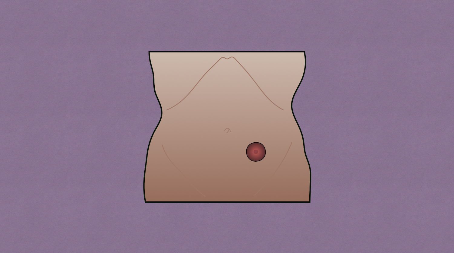
Stomas were intentionally created beginning in the 1700s. Alexis Littré is credited with developing the concept of a need for stoma creation. Another notable development in stoma management was creation of disposable colostomy bags, which were developed by Elise Sørensen after her sister had an ostomy operation in 1953.
Parastomal Hernia
- Occurs when a fascial opening enlarges around the stoma resulting is loops of bowel or intra-abdominal fat protruding above the fascia
- Risk
- Proportion to the time the patient has had the stoma
- Most common after end colostomy
- Least common after loop ileostomy (less pressure)
- Obesity
- Poor muscle tone
- Chronic cough
- Presentation
- Often asymptomatic
- Some patients report change in how appliance fits
- Hernia belt may reduce symptoms
- Treatment
- Asymptomatic → conservative (e.g., stoma belt, weight loss)
- Symptomatic → mesh repair
- Indications: patients with symptoms or inability to adequately pouch the stoma
- Approach
- Primary suture repair → high recurrence rate
- Stoma relocation → if having stoma issues such as narrowing and/or skin excoriation
- Mesh repair
- Keyhole technique → 2-3 cm keyhole cut out of center of mesh; mesh then secured intraperitoneal and secured around bowel to cover the entire fascial defect
- Slit technique
- Sugarbaker technique → bowel secured to anterior/lateral abdominal wall, mesh secured intraperitoneal over bowel loop circumferentially to cover the entire fascial defect
Stoma Prolapse
- Risk factors
- Loop stomas (most common after loop transverse colostomy)
- Obesity
- Parastomal hernia
- Increased intraabdominal pressure
- Pregnancy
- Evaluate for pain, incarceration, bowel ischemia, peristomal skin
- Treatment
- Asymptomatic presentation → hernia belt or support binder
- Can often be reduced with gentle continuous pressure on the stoma while patient is supine
- Applying sugar can help draw out edema and help with reduction
- Surgical indications
- Obstruction
- Ischemia
- Incarceration
Mucocutaneous Separation
- Occurs between the bowel wall and skin
- Must assess for stoma retraction or necrosis, abscess, and fistula
- Prevent early separation by avoiding early use of convexity in pouching system
- Treatment
- Optimize nutrition, smoking cessation, diabetes control, inflammatory bowel disease, and draining infection
- Cleanse area of separation with saline and debride necrotic tissue
- Evaluate wound fully by probing to evaluate for extent of separation
- Fill wound with absorptive product (e.g., hydrofiber, skin barrier powder, barrier paste)
Stoma Necrosis
- Risk factors
- Hypovolemia
- Hypotension
- Edema
- Tension
- Tearing of mesentery
- Presentation
- After surgery, stomas become edematous and often have a change in the mucosa from pink to darker red
- Cyanosis of mucosa or sloughing of mucosa points to necrosis
- Evaluation
- If not already in place, make pouch transparent to allow for easier examination
- Test tube test
- Insert clear test tube into stoma and inspect with flashlight
- Used to determine if necrosis is below the fascia or above
- Can also evaluate with anoscope or flexible sigmoidoscopy
- Treatment
- Necrosis above fascia → observe and reevaluate – can pursue surgery if patient develops infection or sepsis
- Necrosis below fascia → if patient is stable, requires exploration with likely bowel resection and creation of new stoma
- Full thickness necrosis may result in stoma stenosis or mucocutaneous separation
Stoma Retraction
- Stoma ≥0.5 cm below skin surface
- Mechanisms
- Tension → related to obesity or mesenteric edema causing bowel to be pulled down to the level of the skin
- Skin around the bowel sinks in relative to the regular contour of the abdomen causing the stoma to sit in a crease
- Poor initial ostomy site selection or significant weight loss are common causes
- Risk factors
- Obesity
- Tension
- Initial stoma height <1 cm
- Treatment
- Stays above fascia → local wound care
- Retracted below fascia → surgical revision
Stoma Stenosis
- Narrowing of stoma that impairs its normal function – can occur at level of fascia, skin, or both locations
- Presentation
- Cramping followed by episodes of high output
- Bloating
- Pain
- Erratic output
- ± Squirting of stool under pressure
- Evaluate with digital exam to assess for degree of stenosis and location of stenosis (skin and/or fascia)
- Treatment
- Asymptomatic
- Gentle dilation with finger or dilator can provide short-term relief – repeat dilation should be avoided because it can result in scarring and even more stenosis
- Low-residue diet can help relieve symptoms and help with passing stool easier
- Laxatives, stool softeners
- Symptomatic → surgery
- Local revision with skin excision and rematuring
- Consider reseating at different location if skin excoriation is present or a parastomal hernia is present
- Asymptomatic
High Ostomy Output
- Output >1500 cc/day (normal output ranges from 600-1200 cc/day)
- Complication of small bowel ostomies
- Risk factors
- Short bowel
- Sepsis
- Diabetes
- Medications/prokinetics
- Clostridium difficile enteritis
- Opiate withdrawal
- Internal fistula
- Small bowel diverticula
- Treatment
- Fluid resuscitation
- First-line: fiber supplement (e.g., psyllium/metamucil)
- Second-line: antimotility medications
- Loperamide
- Lomotil
- Tincture of opium
- Codeine
- Other options: octreotide, cholestyramine, PPI
