The first angiographic embolization for rectus sheath hematoma was described by Levy in 1980. The technique included transcatheter use of Gelfoam in order to stop arterial bleeding.
Etiology
- Injury to epigastric vessel branches (usually spontaneous)
- Risk factors: female/elderly, anticoagulation
Epidemiology
- Female predominance
- More common in older patients
- Often on anticoagulation at time of diagnosis
- History of trauma or injury to abdominal wall
- Have been associated with pregnancy
Presentation
- Sudden-onset abdominal pain
- Pain worsens with movement
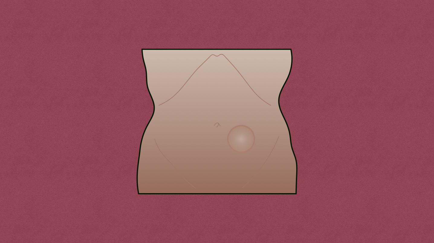
- Abdominal wall mass tender to palpation
- Often have voluntary guarding
- Cullen sign: periumbilical ecchymosis
- Grey Turner sign: blue discoloration in flanks
Imaging
- Ultrasound: heterogeneity in rectus muscle
- CT: high attenuation on unenhanced images
Treatment
- Stable → medical management and observation
- Rest
- Analgesics
- Blood transfusion if needed
- Unstable or enlarging → angiographic embolization
- Surgical evacuation should be considered if skin necrosis, expanding hematoma, or failure of angioembolization occurs
