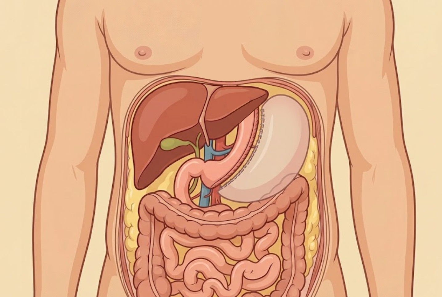
Sleeve gastrectomy is a surgical procedure for weight loss that removes around 80% of the stomach, leaving a tube-shaped stomach behind. The first open sleeve gastrectomy was performed in March 1988 by Dr. Doug Hess in Bowling Green, Ohio as part of a duodenal switch. In 1999, the first attempts were made to complete the surgery laparoscopically by Dr. Michael Gagner.
Indications
- BMI >40 kg/m2
- BMI >35 kg/m2 with significant comorbidities
Contraindications
- Advanced malignancy
- Severe uncontrolled psychiatric disorders
- Uncontrolled GERD
Preoperative Considerations
- Dietary counseling
- Medical clearance → consider EGD or upper GI especially in patients with history of GERD or Barrett esophagus
- Psychological clearance
Relevant Anatomy
- Blood supply
- Pylorus: gastroduodenal artery (from hepatic)
- Lesser curvature
- Left gastric (from celiac)
- Right gastric (from proper hepatic)
- Greater curvature
- Short gastrics (from splenic)
- Left gastroepiploic (from splenic)
- Right gastroepiploic (from GDA)
- Arc of Barklow (anastomosis between right and left gastroepiploic; multiple branches)
- Angle of His: acute angle created between the cardia and the esophagus
- Incisura angularis: sharp angle located in the lesser curvature at the junction of the body and antrum of the stomach
- Prepyloric vein of Mayo: small vein that drains pyloric region of the stomach
Surgical Technique
- Access the abdominal cavity and perform a general inspection of the abdomen. Trocars will be placed to left of midline in supraumbilical region, two auxiliary 5-mm trocars in subxiphoid (for liver retraction), and one along anterior axillary line before rib 12 for assistant. Liver is retracted using fan 2and four additional 12-mm trocars are inserted into RUQ, epigastrium, LUQ, and right paramedian region.
- Pylorus is identified with palpation and visualization of the prepyloric vein of Mayo. The short gastric vessels are divided with ultrasonic energy approximately 2-6 cm from the pylorus in order to allow for entry into the lesser sac. The remaining short gastric vessels are divided by continuing to divide along the greater curvature of the stomach.
- At the level of the proximal body of the stomach, the gastrosplenic ligament is constituted by two separate leaflets (one more anterior and one more posterior). The leaflets can be divided separately if needed depending on their thickness. The splenic artery may also be visualized at this point. The fundus of the stomach is retracted gently due to close proximity to the spleen and the upper body of the stomach is also retracted inferomedially to expose the first short gastric vessels and left crus of diaphragm.
- Hiatal hernia repair may be performed at this time if indicated.
- Posterior gastric adhesions are lysed to allow for complete visualization of the lesser curvature of the stomach. An orogastric tube is placed by anesthesia and advanced to the antrum under visualization. Cephalad and medial retraction of the gastric fundus is necessary at this point as the esophagogastric junction is located posteriorly.
- After bougie is placed, the 60-mm linear stapler is used to sequentially divide the stomach.
- First firing is 2-6 cm from the pylorus and oriented horizontally towards the patient’s left shoulder.
- Careful attention should be given at the level of the incisura angularis to avoid excessive narrowing.
- At the esophagogastric junction, the stapler should be fired without abutting the bougie
- Gastric staple line is then reinforced either with absorbable synthetic or biologic staple line reinforcement strips or by oversewing the staple line in its entirety or in parts.
- Specimen is removed and ports are closed.
Postoperative Considerations
- ERAS protocols
- Usually discharged within 23 hours of procedure
- PRN Zofran plus Prochlorperazine and/or scopolamine patch
- Clear liquid diet on POD1 with discharge home if tolerating
- Upper GI contrast studies can be completed in patients with PO intolerance or clinical derangements from the normal postoperative course
Postoperative Complications
- Bleeding (15%)
- Most common locations
- Devascularization of greater curvature of stomach
- Short gastric vessels
- Spleen
- Port site
- Most common locations
- Leak
- Less likely to close spontaneously compared to RYGB
- Etiology
- Ischemia
- Staple line hematomas
- Staple malfunction
- Increased intraluminal pressure
- Timing
- Acute (<7 days)
- Ischemic leaks are apparent POD5-7
- Early (1-6 weeks)
- Late (>6 weeks)
- Chronic (>12 weeks)
- Acute (<7 days)
- Presentation
- Fever, tachycardia, tachypnea
- Abdominal pain
- Increased WBC and CRP
- Usually occurs in proximal aspect of sleeve at esophagogastric junction as it is the most vulnerable in the presence of distal stenosis
- Diagnosis:
- CT A/P
- Contrast UGI
- Not as sensitive at CT A/P
- Can be false negative if obtained early postop before most leaksk have yet to occur
- Stenosis