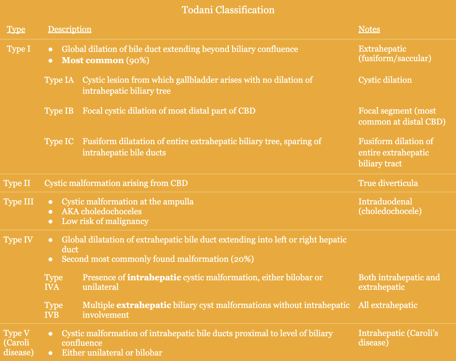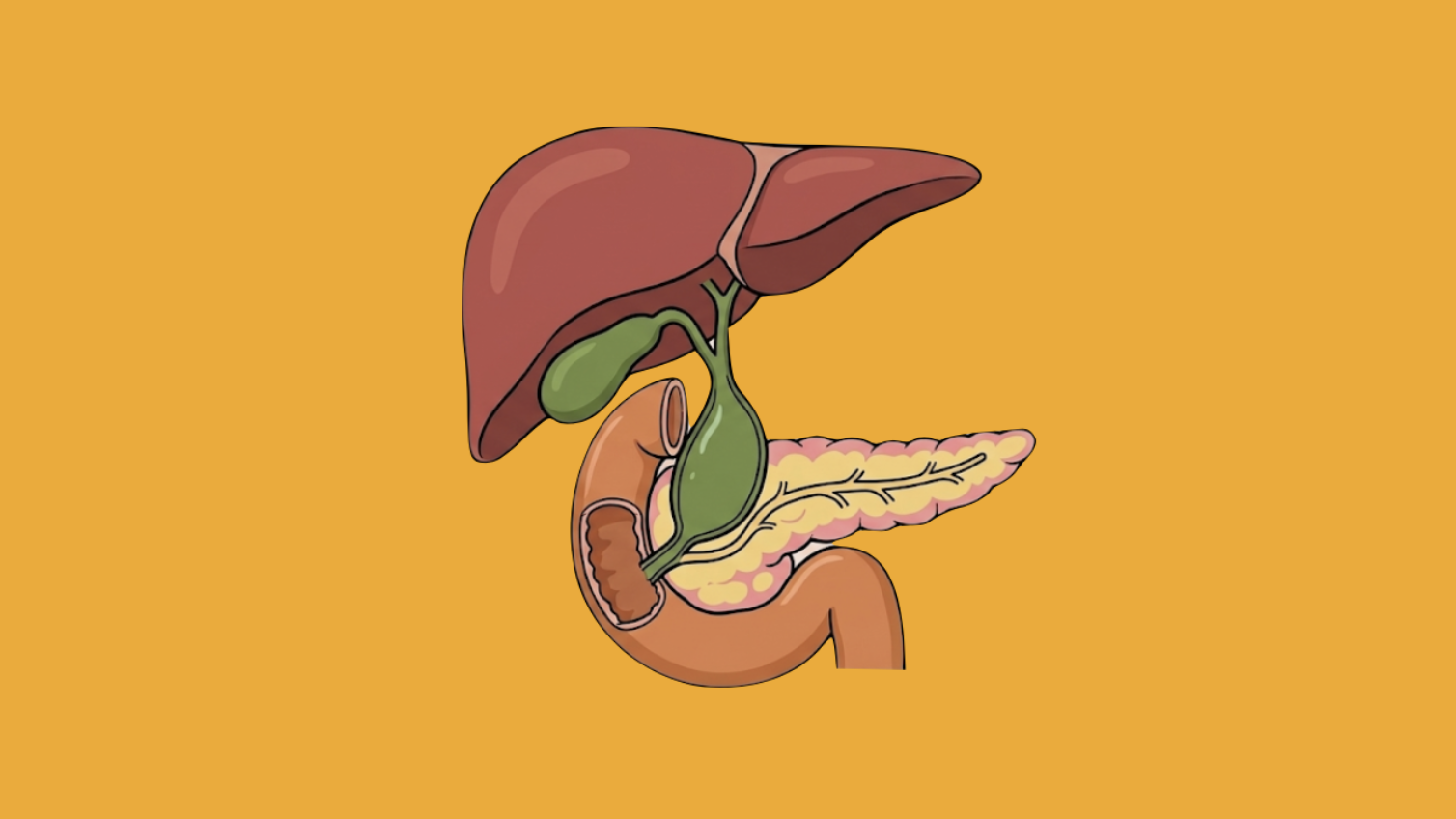
Choledochal cysts are malformations that affect the biliary tract and are often diagnosed in the newborn or antenatal period. There is a malignant transformation risk and this concern is what guides the majority of investigation and treatment decisions. The Todani classification is used to describe the different types of choledochal cysts and was devised in 1977 by Japanese pediatric surgeon Takuji Todani.
Epidemiology
- More common in Asia → Japan and China
- Majority (80%) are diagnosed at birth
- Female predominance (4:1)
Pathogenesis
- Multiple proposed theories
- APBDU (highly accepted)
- Abnormal fused channel between CBD and pancreatic duct → anomalous pancreaticobiliary duct union (APBDU)
- Reflux of pancreatic fluid into biliary tract leading to digestion of bile duct lining
- Congenital malformation
- Obstructive biliary stenosis
- Abnormally high pressure within biliary tree
Presentation
- Classic triad (rare!) → abdominal pain, jaundice, RUQ mass
- Children
- Largely asymptomatic – can have jaundice or mass
- Usually diagnosed on ultrasound
- Adults (rare)
- Abdominal pain secondary to biliary disease which persists after cholecystectomy
- This prompts further workup and eventual diagnosis of choledochal cyst
- May present late with malignancy
Workup
- Abdominal ultrasound
- Limited sensitivity
- Can visualize biliary dilation but doesn’t allow for enough detail
- CT
- Critical in diagnosis and treatment
- Highly sensitive
- Allows for greater detail of surrounding MRI
- Visualize complications related to choledochal cysts → pancreatitis, biliary-free fluid, cholecystitis
- MRCP
- Provides most useful information; preferred
- Allows for cross-sectional imaging of intra- and extrahepatic biliary tree and pancreatic duct
- Visualization of stricturing, obstruction, or evidence of malignancy
- ERCP
- May be utilized if MRI or MRCP leaves uncertainty
- Assess for other causes of biliary dilatation or periampullary masses
Treatment
- Type I → resection and Roux-en-Y hepaticojejunostomy reconstruction
- Resection: CBD through common hepatic duct (divided just distal to confluence) and cholecystectomy
- If adherent to portal vein: anterior excision and posterior mucosectomy
- May require pancreaticoduodenectomy if extending into head of pancreas
- Type II → simple diverticulectomy
- Type III
- <3 cm → ERCP (marsupialization)
- >3 cm → surgery (transduodenal excision and sphincteroplasty)
- Type IV
- IVA → partial liver resection and Roux-en-Y hepaticojejunostomy reconstruction
- Also requires extrahepatic resection similar to Type I/IVB
- Diffuse intrahepatic involvement may prevent excision and thus may require liver transplant
- IVB → resection and Roux-en-Y hepaticojejunostomy reconstruction
- Resection similar to Type I – common bile duct through the common hepatic duct (divided just distal to confluence) and cholecystectomy
- IVA → partial liver resection and Roux-en-Y hepaticojejunostomy reconstruction
- Type V → supportive
- If fails: resection (if able) versus liver transplant