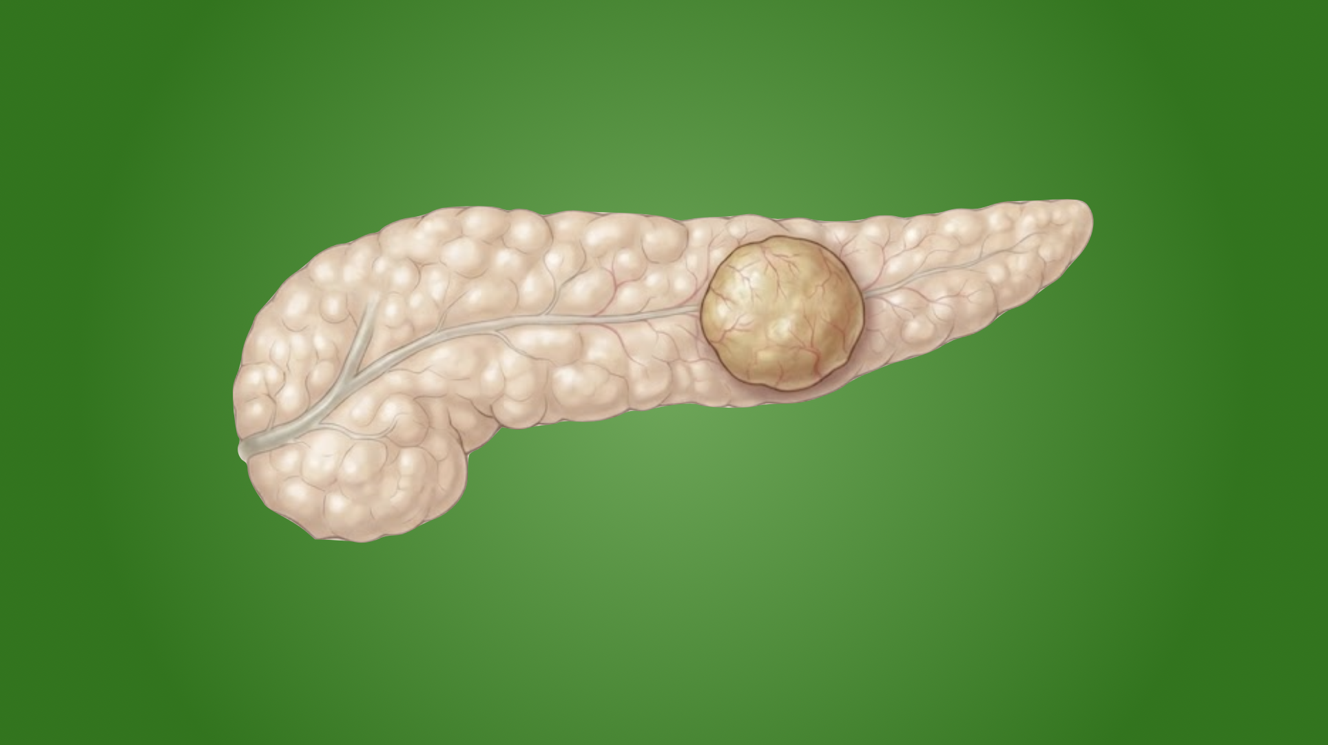
Glucagonoma is a a pancreatic neuroendocrine tumor (PNET) that secretes glucagon, resulting in elevated blood glucose. S. William Becker first described a glucagonoma in 1942 in his paper “Cutaneous Manifestations of Internal Malignant Tumors” in Archives of Dermatology and Syphilology.
Etiology
- Sporadic (most commonly)
- Associated with MEN type 1 (5-17%)
Epidemiology
- Female predominance (2-3:1)
- Average age 52-54
Pathogenesis
- Malignant
- Most are sporadic, rarely have MEN1 or von Hippel-Lindau syndrome
Presentation
- Weight loss (depletion of fat and protein stores, vitamin deficiencies – most common)
- 4Ds
- Diabetes (76-94%)
- Dermatitis
- Deep vein thrombosis
- Depression
- Necrolytic migrating erythema (66%) → painful, bullous dermatosis that evolves into patches with ulceration
- Starts as erythematous papules of face, perineum, and extremities
- Enlarge and coalesce over 1-2 weeks
- Often pruritic
Workup
- Fasting glucagon >500 pg/mL (often >1000)
- Localize with CT or MRI
- Somatostatin receptor imaging
- Consider if initial localization scan fails
- Options
- Somatostatin (octreotide) receptor scintigraphy → classic
- Functional PET scan (Ga-68 DOTATATE)
- If noninvasive fails, can pursue invasive imaging
- Endoscopic ultrasound
- Selective visceral angiography → gold standard but most invasive
Treatment
- Medical management
- Supplemental enteral nutrition
- Octreotide is given to reverse catabolic state
- IV infusion of amino acids to reverse symptoms and improve dermatitis
- Thromboembolism prophylaxis
- Resectable disease should undergo complete anatomic resection
- Head/neck → pancreaticoduodenectomy
- Body/tail → distal pancreatectomy (concurrent splenectomy if malignancy concern)
- Entire pancreas → total pancreatectomy
- Can consider enucleation for small tumors (<2-3 cm), but this is controversial
Relevant Information
- Average size at diagnosis is 5-10 cm
- 65-75% are located in body or tail of pancreas (corresponds to alpha cell location)
- Malignant in 50-80% of cases. 80% of patients have liver metastasis at time of diagnosis
Resources