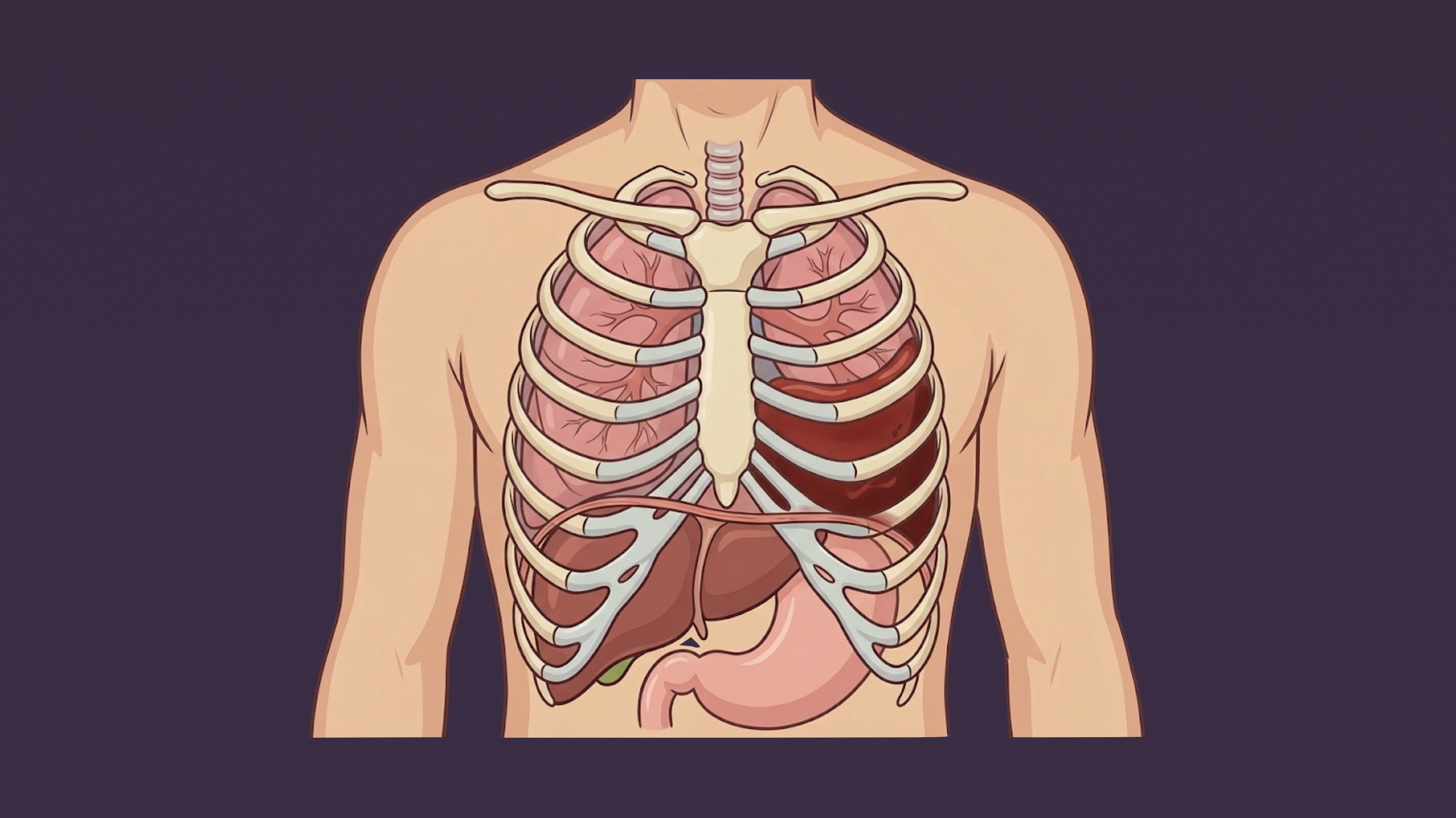
Retained hemothorax refers to blood within the thoracic cavity that has failed to evacuate after chest tube drainage for initial hemothorax for 2-3 days.
Risk Factors
- High initial volume of blood
- High injury severity score
- Mechanical ventilation
- Multiple rib fractures
- Pneumonia
- Prolonged time of chest tube drainage
- Low hematocrit
Presentation
- Small retained hemothorax
- Not clinically relevant
- Parietal pleura will break down and absorb blood
- Larger retained hemothorax
- ~300 mL
- Dullness to percussion
- Egophony: patient says “E” but sounds like nasal “A” through stethoscope
- Tactile fremitus: palpable chest vibrations when patient speaks
- Fine crackles most notable in basilar lung fields
- Persistent oxygen requirement
- Dyspnea
- Shortness of breath
Workup
- CXR: persistent blunt or obscured costophrenic angle
- CT with IV contrast
- Best method for determining volume of retained hemothorax
- IV contrast is important to distinguish fluid from consolidated or contused lung parenchyma
- Determine volume by using formula: v = d2 x l
- d = greatest depth of hemthorax from chest wall
- l = greatest length of hemothorax in cranial-caudal direction
- Ultrasound: good screening modality but not good at quantifying fluid
Treatment
- Initial hemothorax → chest tube insertion followed by CXR to evaluate for tube positioning
- Persistent opacity or poor evacuation of hemothorax after chest tube placement should prompt consideration for risk of retained hemothorax. If present, chest tubes can be either replaced or repositioned with the hemothorax monitored for clearance with daily CXRs.
- If retained hemothorax by hospital day 3, thoracic CT with IV contrast should be obtained to evaluate the volume of retained hemothorax.
- Volume <300 mL → existing chest tube, remove once output <200 mL per day
- Volume ≥300 mL → VATS
Relevant Information
- Retained hemothorax volume >300 mL have been shown to require additional interventions to evacuate.
- There is a higher conversion from VATS to open thoracostomy when retained hemothorax is operated on after hospital day 7
- Treatments second line to VATS for patients who are poor surgical candidates
- Tissue plasminogen activator (t-PA)
- Image-guided drain placement with or without t-PA
Complications
- Empyema
- Fibrothorax
- Formation of located effusions that result in adhesive bands that prevent lung expansion → trapped lung
- Persistent atelectasis, restricted lung function
Resources
- EAST Guidelines: Management of Hemothorax and Occult Pneumothorax
- WEST Guidelines: Management of Hemothorax