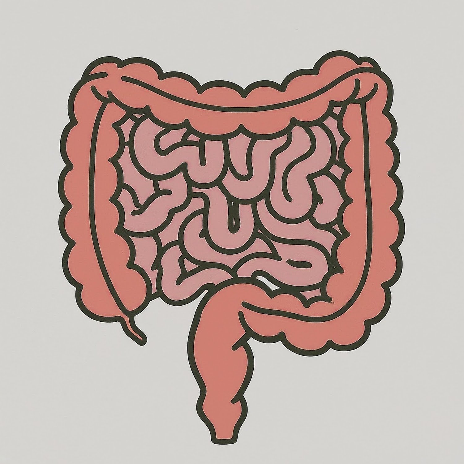
Small bowel obstruction is a blockage of the small intestines. It was recognized as early as the sixth century B.C. by Susruta. From then until recently, intestinal obstruction was recognized only in connection with a strangulated hernia. Owen Wangensteen focused on intestinal obstruction in 1942 and stated that J.A. Hartwell and J.P. Hoguent had conducted the first important experimental study on intestinal obstruction in 1912. Hartwell and Hoguent had worked with dogs, causing them to have intestinal obstructions, and then treating them with large amounts of subcutaneous saline.
Etiology
- Postoperative adhesions (most common cause in the U.S.)
- Congenital anatomic abnormalities
- Midgut volvulus
- Ileal atresia
- Adhesions
- Extraluminal causes
- Adhesions
- Hernias
- Carcinomas
- Abscesses
- Intrinsic to bowel wall: primary tumors
- Intraluminal causes
- Gallstones
- Enteroliths
- Foreign bodies
- Bezoars
- Radiation enteropathy
Presentation
- Nausea and vomiting
- PO intolerance
- Abdominal pain, colicky in nature
- Abdominal distention
- Obstipation
- Localized tenderness, rebound, guarding → suggests peritonitis (possible strangulation and ischemia)
- Dehydrated appearance
- Tachycardia
- ± Hypotension, rare
- ± Shock, rare
- ± Fever → suggests possible strangulation
Workup
- BMP, Mg, P, CBC with differential, and lactate
- Lactic acidosis → suggests intestinal ischemia/necrosis
- Leukocytosis → suggests strangulation
- Oliguria: urinary output <400 mL/day OR <20 mL/hr
- Abdominal X-ray
- Initial imaging of choice
- Dilated gas or fluid-filled loops of small bowel, gasless or nondistended colon
- May demonstrate the cause of obstruction (e.g., foreign body, gallstones)
- CT A/P w IV contrast
- 95% sensitive and specific – less sensitive in patients with partial SBO
- PO contrast may not be used on initial CT due to delay and concern for aspiration
- Dilated bowel loop >2.5 cm is concerning for high-grade SBO
- Identify transition point in 93% of cases
- Findings concerning for bowel wall compromise
- Bowel wall edema or hemorrhage
- Altered bowel wall enhancement
- Interloop ascites
- Mesenteric edema/fat stranding
- Vascular engorgement
- Vessel occlusion
- Pneumatosis
- Barium studies
Treatment
- Nonoperative management
- IV fluid resuscitation
- NG tube decompression
- Based on old, debated evidence
- Empties stomach and reduces risk of aspiration
- Contrast challenge with gastrografin
- Diagnostic and therapeutic
- Gastrografin in the colon in 24 hours predicts resolution without surgery
- Surgery
- Considered after nonoperative management is attempted for 2-3 days and is unsuccessful or if evidence of peritonitis or clinical deterioration
- Etiology determines surgical intervention
- Adhesions: lysis of adhesions
- Hernia: manual reduction and closure of defect
- Malignant tumor: nonoperative management; intestinal bypass of obstructing lesion
- Intraabdominal abscess: percutaneous drainage of abscess; laparotomy; laparoscopy
- Special situations
- Closed-loop small bowel obstruction has an increased risk for strangulation and failure of nonoperative management
- Small bowel obstruction with transition point at hernia site may require urgent surgery if ischemia bowel is suspected
- Pregnancy is not absolute contraindication to CT scans and overall management is the same
Relevant Information
- SBOs make up 80% of all bowel obstructions.
- Third spacing occurs when water and electrolytes accumulate intraluminally and in the bowel, which also accounts for the dehydration and hypovolemia
- Flora of the small intestine changes with obstruction. Escherichia coli, Streptococcus faecalis, and Klebsiella spp. are most common.
- A proper physical exam should be conducted to rule out incarcerated hernias in the groin, femoral triangle, and obturator foramen
- Patients who have required operation in the past for SBO have roughly 15% incidence of operative intervention
Classifications
- Partial small bowel obstruction (pSBO): incomplete obstruction with luminal narrowing
- Complete small bowel obstruction: obstruction with no passage of luminal contents
- Complete small bowel obstruction with bowel compromise: ischemia/injury leading to bowel necrosis and/or bowel wall perforation