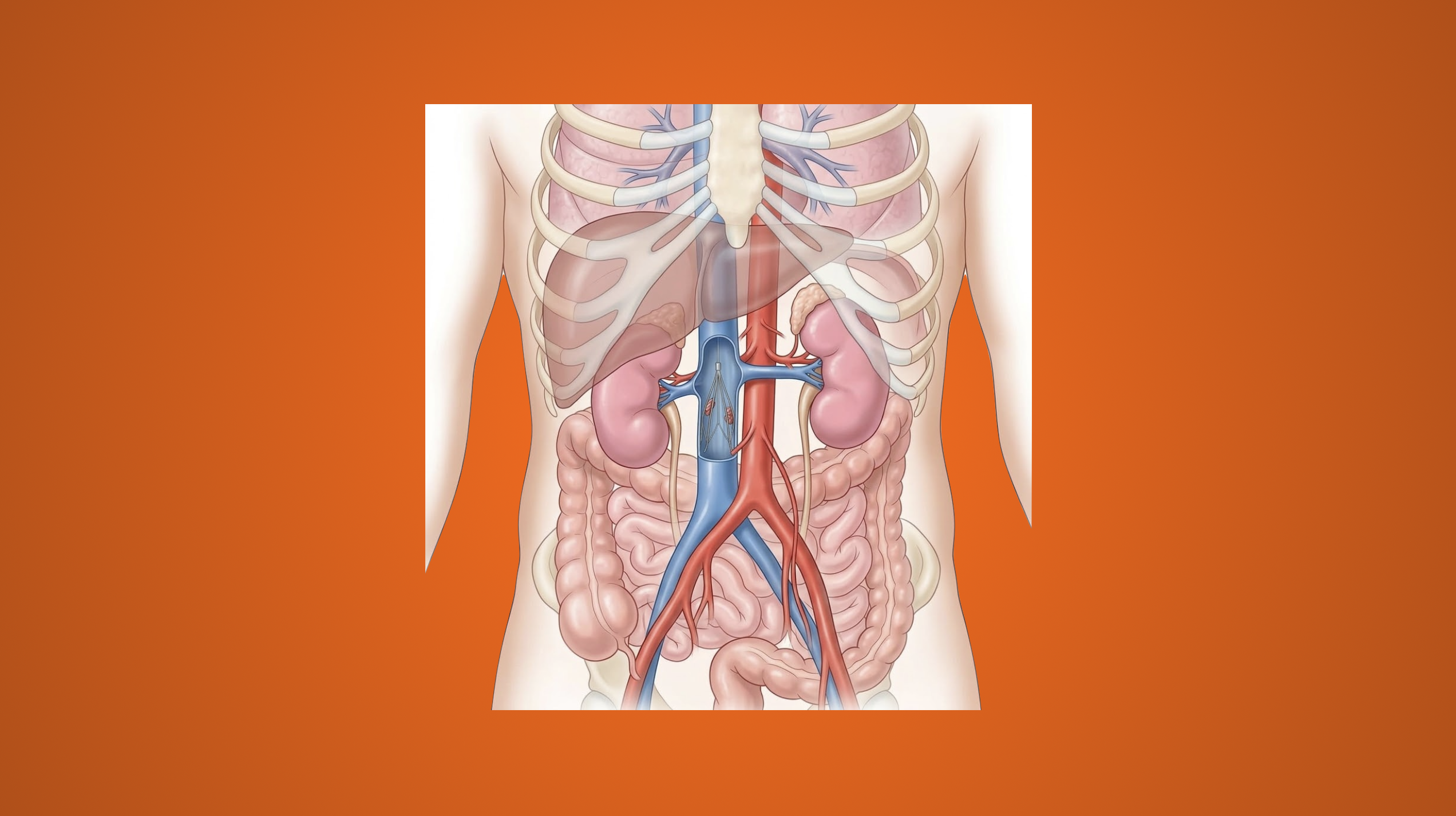
Vena cava filter placement is indicated in patients with venous thromboembolism (VTE) who either have contraindications to anticoagulation or have failed treatment. The goal of these filters is to prevent pulmonary embolism (PE) by trapping the emboli in the device.
The first filter was introduced by Marion DeWeese in 1958 as a hard-grip filter with silk sutures. Bertman Cohn made and patented an intraluminal filter in 1967. The first transvenous filters was introduced by Kazi Mobin-Uddin around 1968 by making a small venotomy.
Guidelines and Indications
- Guidelines for VTE and IVC filter placement
- Society for Interventional Radiology (SIR, October 2020)
- The Eastern Association for the Surgery of Trauma (EAST)
- VTE: Role of Vena Cava Filter (update in process)
- VTE: Role of Vena Cava Filter (2002)
- Western Trauma Association (WTA)
- VTE Prophylaxis (flowsheet)
- VTE Prophylaxis (entire document)
- American College of Chest Physicians (ACCP)
- Indications
- Absolute indications
- Presence of acute PE or proximal DVT with contraindication to anticoagulation
- Progressive venous thromboembolic disease despite adequate anticoagulation
- Significant bleeding complications while on anticoagulation for VTE
- Inability to maintain adequate anticoagulation
- Relative indications
- Free-floating caval or iliofemoral vein thrombus (≥6 cm in length)
- Pulmonary thromboembolectomy or thrombolytic therapy of iliocaval thrombus
- Poor medication compliance
- Significant fall risk
- Acute PE with poor cardiopulmonary reserve
- Prophylactic indications → no evidence of thromboembolic disease but patient is at high risk for development
- Potential patients: severe trauma, closed head injury, spinal cord injury, multiple long-bone or pelvic fractures, high-risk immobilized patient
- OFF-LABEL, NOT APPROVED BY FDA
- Absolute indications
- Relative contraindications
- Nickel hypersensitivity
- Bacteremia
- Severe coagulopathy
- Anticoagulation should be resumed when contraindication has resolved
Types of Vena Cava Filters
- Permanent IVC filters → no mechanism for removal
- Optional (retrievable and convertible) IVC filters → can be removed or left long-term
- Convertible IVC filters → may be deactivated by percutaneous conversion by changing the filter to an open configuration
Filter Location
- Infrarenal IVC is generally the most preferred location as it minimizes the risk of obstructing the renal veins.
- Suprarenal Indications
- Compromised infrarenal IVC
- Pregnancy
- Gonadal or renal vein thrombosis
- Thrombus extending to level of renal veins
- Significant infrarenal thrombus burden
- Anatomic variants
- Superior vena cava (SVC) placement
- Ideally placed immediately proximal to innominate vein
- Orientation of filter should be reversed compared to IVC filter
- Femoral IVC filter kit used for jugular approach
- Jugular IVC filter kit used for femoral approach
- Filter position confirmed with CXR
Filter Placement
- Preoperative planning
- CT or MRI A/P → evaluate venous anatomy and any anomalies
- Bilateral lower extremity venous duplex ultrasound → confirm site and extent of DVT
- Options for managing anomalies
- Duplicate IVC
- Place suprarenal
- Place filter in each IVC
- Isolated left-sided IVC
- Common femoral vein access is preferred
- May require suprarenal placement
- Circumaortic and retroaortic left renal veins
- Types
- Type I: single renal vein bifurcating into two separate veins
- Type II: two left renal veins
- Type III: multiple pre- or retroaortic veins
- Place filter inferior to entire circumaortic venous complex
- Place between lowest renal vein and iliac confluence in suprarenal IVC
- Types
- Megacava
- IVC measuring >28 mm in diameter
- Place Bird’s Nest IVC filter as it can be placed in IVC diameters >32 mm
- Place IVC filers in bilateral iliac veins
- Duplicate IVC
- Fluoroscopy-guided IVC filter placement
- Right common femoral vein or right internal jugular vein are preferred sites as they provide a straight course to the IVC
- Venogram is performed to evaluate the anatomy and exclude anatomic variants. IVC is measured, distance between lowest renal vein and iliac confluence is measured, and no thrombus in IVC is noted.
- Most IVC filters are placed below the level of the renal veins to avoid future renal dysfunction if the IVC filter were to thrombose
- Filter is deployed and position is confirmed. Filter is unsheathed. Completion venography performed. Completion KUB ordered to document filter position for future reference
- Ultrasound-guided IVC filter placement (IVUS)
- May be utilized in patients who are too unstable for transfer to fluoroscopy suite
- Venous access options
- Double femoral venous puncture → right common femoral vein preferred
- Single puncture, single sheath → no real-time imaging of IVC filter positioning and deployment
- Pullback technique allows for landmark identification (liver, hepatic veins, renal artery, origins of renal veins, iliac vein confluence) and IVUS catheter placed at lowest renal vein. IVC filter delivery sheath is advanced over-the-wire. Constrained filter is inserted into delivery sheath and positioned at distal end of sheath which is confirmed with IVUS imaging.
- Filter is deployed in a “pin and pull” manner and IVUS catheter is carefully removed. IVC filter position confirmed with KUB.
Filter Removal
- Should be performed when contraindication to anticoagulation has resolved
- Benefits of removal
- Decreased risk of long-term implantation
- Decreased risk of subsequent DVT
- No filter migration/embolization
- IVC stenosis or occlusion
- Timing of removal
- Optional IVC filters → remove via jugular approach after performing venogram of IVC to ensure no thrombus either in IVC or filter itself
- Prophylactic filter → remove 29-54 days after placement (risk of PE has passed, risk/benefit profile favors removal)
Filter Complications
- Early complications
- Access site thrombosis (8.5%)
- Filter tilt
- Angulation >15º from long axis of filter → results in decreased efficacy and increased risk of recurrent PE
- Should be addressed during initial procedure if possible
- Risk increases with placement of conical IVC filter
- Most common cause of retrieval failure
- Malposition (1.3%)
- Hematoma (0.6%)
- Arteriovenous fistula (0.02%)
- Late complications
- Filter penetration
- Associated with increased dwell time and degree of filter tilt
- May lead to pseudoaneurysm, bowel penetration and injury, infection, and pain
- Grade
- Grade 0: normal, filter strut confined entirely within IVC
- Grade 1: filter strut is immediately adjacent to external aspect of IVC wall likely reflecting tenting of IVC wall
- Grade 2: filter strut is entirely outside IVC lumen within retroperitoneum as evidenced by “halo” of retroperitoneal fat around axially viewed strut
- Grade 3: filter strut interacts with adjacent organ outside of IVC
- Remove filter is patient is symptomatic
- Filter fracture
- Most common late complication of retrievable filters
- Loss of structural integrity of the device
- Associated with increased dwell time and mechanical stress
- Increased risk of embolization and migration into hepatic veins, right heart, and pulmonary arteries
- Filter migration
- Change in craniocaudal position of filter >20 mm or greater than or equal to height of one vertebral body
- Usually occurs within first few months after placement
- Should be removed if it is dislodged
- IVC thrombosis
- Higher in cylindrical filters or filters with umbrella component
- 50% of patients remain asymptomatic
- Treat with systemic anticoagulation
- Filter penetration
Resources
- PREPIC (Prévention du Risque d’Embolie Pulmonaire par Interruption Cave) study
- Long-term, large (n = 400), randomized controlled trial
- Determined safety and efficacy of IVC filters in patients with proximal DVT for prevention of PE
- PREPIC2 trial
- Randomized, open-label trail
- Assigned hospitalized patients with severe acute PE to retrievable IVC filter implantation plus anticoagulation (n = 200) versus anticoagulation alone with no filter placement (n = 199)
- PRESERVE (Predicting the Safety and Effectiveness of Inferior Vena Cava Filters) study
- Collaboration between Society for Vascular Surgery, Society of Interventional Radiology, and U.S. Food and Drug Administration
- Evaluate safety, effectiveness, and current patterns in use of IVC filters
- Enrolled 2,100 patients at 60 centers in the United States
- Results to be determined