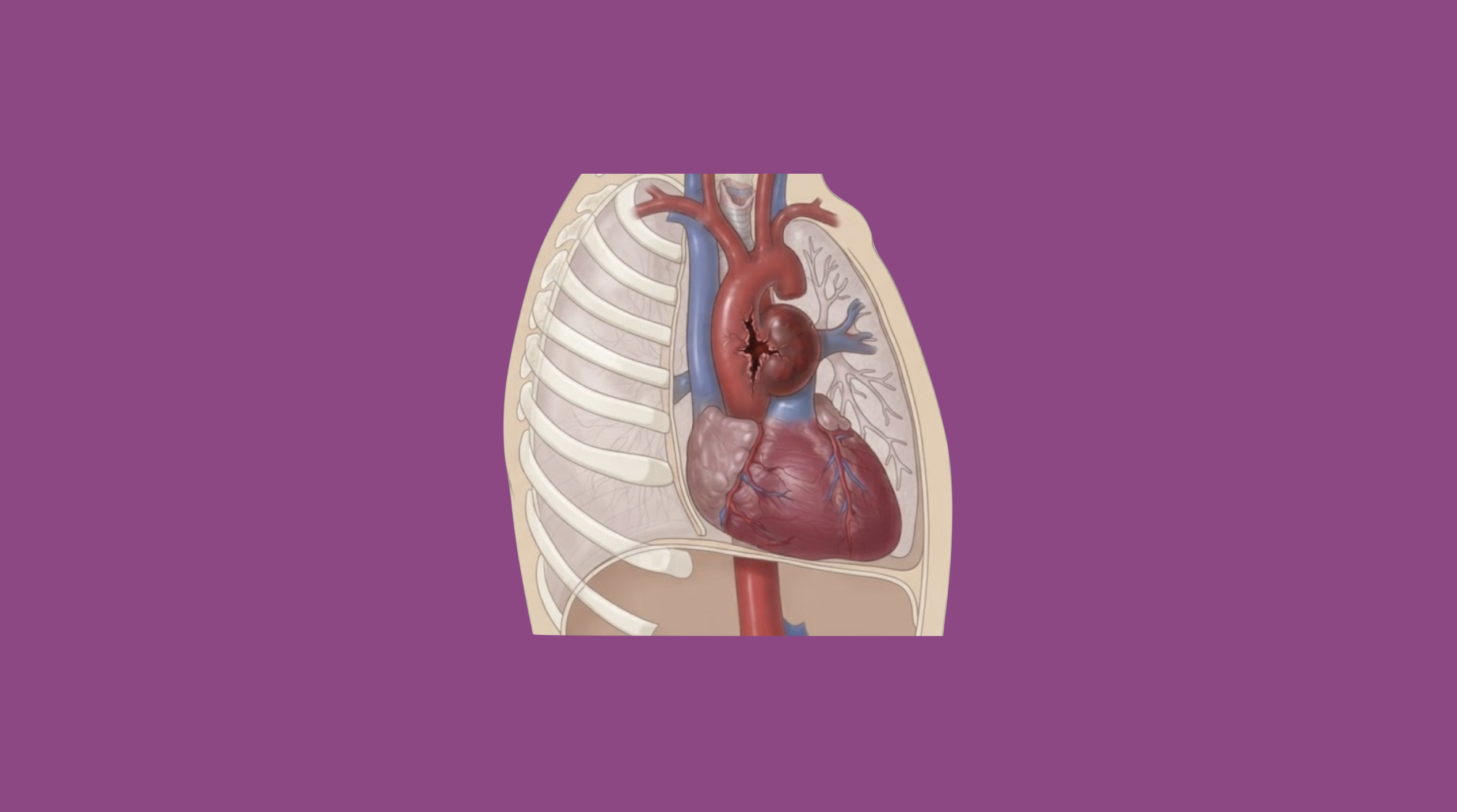
Blunt thoracic aortic injury (BTAI) occurs as damage to the aorta secondary to blunt mechanism, often high-impact motor vehicle collision. In 1958, Dr. Parmley published a review after investigating 275 cases of non-penetrating aortic injuries. At that time, only 14% of patients reached the hospital alive. Since then, medicine has evolved and we now have multiple treatment modalities to help improve outcomes.
Etiology
- High-impact trauma
- Motor vehicle collisions (most common, 60%)
- Motorcycle accidents
- Pedestrians struck by automobiles
- Fall from significant heights
- Crush injuries
Epidemiology
- Median age: 41
- Male predominance
Pathogenesis
- Rapid deceleration causing shear stress and tearing of the aortic wall
- Most common location is proximal descending thoracic aorta (at aortic isthmus) as it is the transition point between the mobile arch of the aorta and the fixed descending thoracic aorta
Presentation
- May not have obvious signs of injury
- Unconscious
- Normal vital signs including blood pressure do not rule out BTAI
- Chest wall ecchymosis, crepitus
- Flail chest
- Sternal fracture
- Absent breath sounds
- Unequal pulse exam
Workup
- CXR
- Widened mediastinum (>8 cm)
- Abnormal aortic contour
- Tracheal deviation
- Apical capping
- Depressed left mainstem bronchus
- Left hemothorax
- CTA C/A/P → gold standard
- Mediastinal or periaortic hematoma
- Intimal flap
- Important for diagnosis as well as preoperative planning
- Requires high index of suspicion
BTAI Classification
- Proposed by Azizzadeh et al. and adopted by Society for Vascular Surgery
- Uses imaging to determine extent of injury to the anatomic layers of the aorta
- Grades
- Grade 1: isolated intimal tear (25%)
- Grade 2: intramural hematoma (15%)
- Grade 3: pseudoaneurysm (50%)
- Grade 4: frank aortic rupture (10%)
Treatment
- ATLS protocol and stabilization
- Medical management
- Anti-impulse therapy
- Reduces risk of rupture (from 12% to 1.5% per one study)
- No consensus on target blood pressure or heart rate
- SBP <120 mmHg
- HR <60
- Can utilize Esmolol drip as it is effective and can be titrated
- Admit to ICU
- Place arterial line
- Anti-impulse therapy
- Recommendations
- Grade 1 → management with serial imaging
- Grade 2-4 → urgent (<24 hours) intervention with TEVAR
- Thoracic endovascular aortic repair (TEVAR) is preferred
- Requires access vessel 7 mm in diameter free of calcification and tortuosity
- Proximal and distal landing zones must include 2 cm of healthy aorta for adequate seal
- Coverage of the left subclavian artery may be necessary to obtain an adequate proximal seal. Contraindications to coverage of left subclavian artery
- Existing coronary artery bypass graft with left internal mammary artery
- Functional left AVF or AVG
- Dominant left vertebral artery
- Left vertebral artery terminating in posterior inferior cerebellar artery
- Postoperative care
- Anti-impulse therapy is no longer needed – hemodynamic parameters can tailor to other injuries
- Physical exam including assessment of bilateral groin access sites (hematoma, pseudoaneurysm), distal extremity pulse exam (ischemia due to thrombosis or embolism), neurologic exam to monitor for SCI and/or stroke
- Follow up for imaging surveillance using CTA C/A/P at 1 month, 6 months, and annually thereafter
Relevant Information
- Aortic zones
- Zone I: left subclavian to celiac trunk
- Zone II: celiac trunk to renal arteries
- Zone III: renal arteries to bifurcation
- Majority occur at aortic isthmus, distal to the left subclavian artery. Coverage of the left subclavian artery may be necessary to obtain an adequate proximal seal
- Aortic-related mortality is relatively rare in patients with Grade 1-3 BTAI