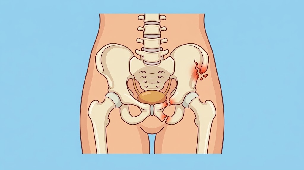
Pelvic fractures are estimated to have an incidence of 27-37 per 100,000 patients each year. They are more frequently seen in patients who present with polytrauma and are usually the result of a trauma with a high mechanism.
Etiology
- Motor vehicle and motorcycle collisions (40-60%)
- Pedestrian versus automobile collisions (20%)
- Falls (5-30%)
Anatomy
- Pelvis
- Bony ring formed by two innominate bones, sacrum, and coccyx
- Retroperitoneal zones
- Zone 1
- Central retroperitoneum – diaphragm to aortic bifurcation
- Contains abdominal aorta, IVC, portions of duodenum, and pancreas
- Zone 2
- Bilateral perinephric regions, from renal vasculature medially to white line of Toldt lateral and from diaphragm superiorly to aortic bifurcation inferiorly
- Contains kidneys, renal vasculature, adrenal gland, and ureter
- Zone 3
- Inferior to aortic bifurcation
- Contains common, internal, and external iliac arteries and veins, distal ureters, and distal sigmoid colon and rectum
- Zone 1
Injury Classification
- Young-Burgess System
- Characterizes pelvic fractures
- Lateral compression (LC) injuries
- Most common pelvic injury
- Classically occur after T-bone motor vehicle, pedestrian versus automobile collisions, and falls from significant height
- Associated with lethal thoracoabdominal trauma and cervical spine injuries
- Must consider missed extrapelvic source of hemorrhage if patient demonstrates hemodynamic instability and/or significant transfusion requirement
- Subtypes
- LC1
- Sacral compression fracture on side of blunt impact with unilateral or bilateral pubic rami fractures
- Most common type of LC injury
- Nonoperative management
- LC2
- Crescent fracture on side of impact
- Often require iliac wing fixation
- LC3
- Applied force extends to opposite hemipelvis to create open book fracture on opposite side of impact1
- Managed individually
- LC1
- Anterior-posterior compression (APC) injuries
- Second most common observed pelvic injury
- Occurs following direct compressive force applied to pelvis or transmitted from femurs, leading to symphysis diastasis and widened pelvic diameter
- Subtypes
- APC I
- Minor widening of symphysis and sacroiliac joints anteriorly, usually <2 cm
- Intact posterior pelvic ligaments
- Don’t require surgery
- APC II
- Widening of anterior sacroiliac joints due to associated disruption of anterior sacroiliac, sacrotuberous, and sacrospinous ligaments
- Intact posterior pelvic ligaments
- Requires treatment with plating or screw fixation of disrupted symphysis
- APC III
- Complete sacroiliac disruption
- Injury to posterior pelvic ligaments
- Requires both anterior and posterior fixation with combination of plating and/or screw placement
- APC I
- Vertical shear (VS) injuries
- Occur after jump or fall from significant height when patient lands one extended lower extremity
- Disruption of all ligamentous attachments of one hemipelvis and subsequent superior and/or posterior displacement
- Managed similarly to APC III injuries
- Open pelvic fractures
- Associated with GI and GU injuries
- 45% mortality rate – most commonly due to early exsanguination due to no tamponade
- Pelvic binder should be placed to aid in underlying hemorrhage
- Peripheral vascular exam should be completed. CTA aorta with bilateral iliofemoral runoff should be completed for discrepancies
Imaging
- Pelvic XR
- FAST
- Completed as adjunct to primary survey in hemodynamically unstable blunt trauma patients
- Can detect 200 mL of intraperitoneal free fluid, but there are several limitations including inability to detect retroperitoneal bleeding
- CT abdomen/pelvis
- Gold standard for diagnosis and characterization of pelvic fractures and retroperitoneal injuries
- May prevent nontherapeutic laparotomy in stable patient with equivocal bleeding source
- Limited to hemodynamically stable patients
Concurrent Injuries
- Thoracoabdominal injury
- Significant association between complex pelvic fractures and blunt aortic injuries
- Low threshold for CTA chest
- Traumatic brain injury (TBI)
- 17% have concomitant TBI
- Abnormal initial neurologic exam requires CT head
- Hypotension and hypoxemia worsen TBI outcomes due to secondary brain injury
- Genitourinary injury
- 16% have concomitant genitourinary injury
- Hematuria is common clinical manifestation
- Retrograde urethrogram (RUG)
- Perform in patients with findings concerning for urethral trauma → blood at urethral meatus, high-riding prostate, scrotal hematoma
- Evaluates urethra
- CT cystography → evaluates bladder
- Vaginal exam is necessary in females
- Management
- Most urethral injuries are managed with diverting subrapubic cystostomy
- Intraperitoneal bladder rupture is repaired in two layer fashion with absorbable suture and catheter decompression
- Rectal injury
- DRE to assess for blood or palpable bone fragments
- Evaluation through either exam under anesthesia with rigid or flexible proctoscopy or sigmoidoscopy may be required
- Neurologic injury
- Lumbosacral nerve injuries are common
- L5 and S1 nerve roots are most often affected
Multimodal Therapy
- Temporary hemostasis
- External compression devices → decrease volume of pelvis and tamponade underlying venous bleeding to restore hemodynamic stability
- Pelvic binders → standard bed sheet or true pelvic binder for stabilization and possible hemostasis, obtain radiograph after placement to ensure adequate pelvic volume reduction, should be taken down every 6 hours to assess for skin breakdown and shouldn’t remain in place >24 hours due to risk of soft-tissue injury and ischemia
- Resuscitative endovascular balloon occlusion of the aorta (REBOA)
- Control presumed and confirmed infradiaphragmatic hemorrhage in hemodynamically unstable patients
- Proper occlusion balloon placement falls into three zones
- Zone I: between takeoff of left subclavian artery and celiac axis; indicated for severe hemodynamic instability or in cardiac arrest in place of resuscitative thoracotomy
- Zone II: between celiac axis and inferior-most renal artery
- Zone III: between inferior-most renal artery and aortic bifurcation; indicated for known pelvic injury
- Preperitoneal packing
- Tamponade venous and bony hemorrhage
- Doesn’t adequately assist with arterial hemorrhage
- Performed through Pfannenstiel or separate lower midline laparotomy. Fascia incised and space of Retzius accessed to open preperitoneal space. Bladder is retracted posterolaterally and ipsilateral retroperitoneal space is bluntly dissected and rapid packing is performed. Six pads usually complete the packing. Fascia is closed to maximize tamponade effect.
- External fixation
- Definitive hemostasis
- Angioembolization
- Goal is to slow bleeding to allow for natural hemostatic mechanisms to take over
- Selective angioembolization
- Risk of inadequate hemostasis
- Gelfoam is agent of choice → remains for several weeks before being absorbed at which point target vessel may recanalize
- Coils → used more often in larger arteries not amenable to Gelfoam
- Coil blockade → microcoils placed in intact distal segment of injured vessel and Gelfoam used to embolize proximal portion
- Open vascular control
- External fixation
- Angioembolization
- Approach to retroperitoneal injuries
- Zone I → require exploration regardless of mechanism; high likelihood of significant underlying vascular injury
- Penetrating zone II and III → exploration
- Blunt zone II and III → observation if no hard signs
Complications
- Pelvic sepsis
- Deep space infections after pelvic trauma/surgery
- Fever, hemodynamic changes, new or worsening leukocytosis, worsening pelvic pain
- Tx: percutaneous drainage of abscesses and antibiotics
- Venous thromboembolism
- Fat embolism syndrome
- May occur after complex pelvic fracture
- Upper body petechial rash, hypoxemic respiratory failure, variable levels of neurologic dysfunction
- Fever, tachycardia, tachypnea, acute kidney injury, anemia
- Cerebral MRI is most sensitive imaging