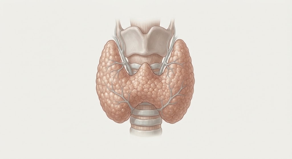
Total thyroidectomy is the surgical removal of the entire thyroid lobe and isthmus. Total thyroid lobectomy refers to removing an entire thyroid lobe and isthmus. The earliest recorded removal of thyroid gland was in 952 AD by Albucasis in order to treat a large goiter. In modern times, it wasn’t until 1971 that Pierre-Joseph Desault performed the first partial thyroidectomy. Later on, Theodor Kocher (1841-1917) introduced more meticulous surgical dissection with using the now-common low collar neck incision, which ultimately won him the Nobel Prize in Physiology or Medicine in 1909.
Indications
- Total thyroidectomy
- Graves’ disease
- Most thyroid cancers (not including low-risk papillary thyroid cancer)
- Multinodular goiter
- Thyroid lobectomy
- Thyroid nodule with uncertain diagnosis
- Symptomatic thyroid nodule
- Low-risk papillary thyroid cancer
Preoperative Considerations
- Thyroid function test
- Neck ultrasound
- Fine needle biopsy
- Molecular testing
- CT neck/chest
- Flexible laryngoscopy → perform in patients with hoarseness or prior neck surgery to evaluate function of RLNs and assess baseline vocal cord function
Relevant Anatomy
- Thyroid gland
- Right and left lobes
- Isthmus: central portion, connects lobes
- Pyramidal lobe (Lalouette’s pyramid): remnant thyroglossal duct
- Ligament of Berry (posterior suspensory ligament)
- Suspends thyroid from the trachea
- VERY close to recurrent laryngeal nerve
- Most common location for recurrent laryngeal nerve injury
- Parathyroid gland
- Superior parathyroid glands
- Located posterior to superior thyroid lobe and RLN
- Inferior parathyroid glands
- Location more variable compared to superior parathyroid glands – often anterior to inferior thyroid gland and RLN or on inferolateral aspect of lobes
- Superior parathyroid glands
- Musculature
- Midline raphe: fibrous seam or ridge formed where muscles or tissues from the left and right sides of the neck join together
- Sternothyroid and sternohyoid muscles cover the anterolateral surfaces of the thyroid gland
- Blood supply
- Superior thyroid artery
- Arise from external carotid artery (first branch)
- Near superior laryngeal nerve
- Inferior thyroid artery
- Arise from thyrocervical trunk
- Near recurrent laryngeal nerve
- Anterior to right RLN
- Posterior to left RLN
- Superior thyroid artery
- Venous drainage
- Superior thyroid vein
- Runs with superior thyroid artery
- Drains into internal jugular vein
- Middle thyroid vein
- Drains into internal jugular vein
- Inferior thyroid vein
- Drains into brachiocephalic (innominate) vein
- Superior thyroid vein
- Nerves
- Recurrent laryngeal nerve (RLN)
- Left
- Arises from vagus nerve at aortic arch
- Loops around ligamentum arteriosum
- Right
- Loops around right subclavian artery
- Innervates intrinsic laryngeal muscles (except cricothyroid)
- Left
- Superior laryngeal nerve
- Most common nerve injury
- Most commonly injured at Ligament of Berry
- Injury results in inability to reach high pitches, difficulty projecting
- Internal branch
- Sensory nerve for supraglottic larynx
- External branch
- Lies on inferior pharyngeal constrictor muscle
- Can be seen during dissection of superior pole of thyroid
- Descends along superior thyroid vessels
- Innervates cricothyroid
- Most common nerve injury
- Recurrent laryngeal nerve (RLN)
Surgical Technique
- Low collar incision created at midline above sternal notch and below cricoid cartilage in a natural skin fold. Dissection carried out through subcutaneous tissue and platysma, creating subplatysmal flaps to enhance exposure. Flaps extend to thyroid cartilage superiorly and suprasternal notch inferiorly.
- Midline raphe is opened and dissection is continued down to the level of thyroid isthmus. Sternohyoid and sternothyroid muscles are dissected and retracted laterally (can be divided if needed).
- Thyroid lobe is retracted medially with finger and space between the thyroid and carotid sheath is opened bluntly until prevertebral fascia is seen. Middle thyroid veins are ligated once exposed.
- Isthmus may be mobilized to further allow dissection. Superior sensory ligament and vessels are divided. The pyramidal lobe can be divided if present. The superior pole of thyroid is retracted and dissection carried carefully especially at this point as external branch of SLN courses through. Superior pole vessels are divided close to the thyroid gland to prevent incidental injury to nerves.
- Lateral and inferior thyroid lobes are dissected after completion of the superior pole. Thyroid lobe retracted medially and elevated anteriorly off paraesophageal space, with identification and preservation of RLN during careful dissection.
- Ligament of Berry is divided, as close to trachea as possible.
- Superior and inferior parathyroid glands are identified and preserved.
- If performing total thyroidectomy, process is repeated on contralateral side.
- Hemostasis is ensured. Strap muscles are reapproximated with absorbable interrupted suture. Platysma is reapproximated with buried interrupted absorbable suture. Skin is closed with subcuticular running suture and skin glue applied.
Postoperative Complications
- Hematoma
- Hypocalcemia (completion thyroid lobectomy)
- Hoarseness
- Vocal cord paralysis
- Wound infection