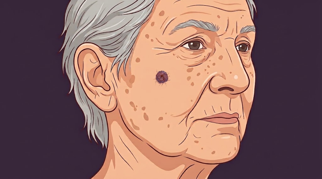
Merkel cell carcinoma is a type of skin cancer that stains positive for CK20 and several neuroendocrine markers. On physical exam, it appears as a firm, painless red lesion. Its name after Friedrich Sigmund Merkel, a German anatomist who first described its appearance under microscopy in 1875. Prior to this, it was called “trabecular carcinoma” by Cyril Toker in 1972.
Etiology
- Risk factors: UV radiation, sun-exposed skin, immunosuppressed, solid organ transplant, HIV
Epidemiology
- Fair skin
- Male predominance (2:1)
- Associated with other malignancies
- Multiple myeloma
- Chronic lymphocytic leukemia
Pathogenesis
- Merkel cell polyomavirus infection
- Not virally associated → mutation pattern is consistent with UV radiation
Presentation
- Firm, red lesion
- Grows fairly rapid, often ulcerates
- Painless, nontender
Workup
- Biopsy
- CK20: characteristic paranuclear dot-like or diffuse cytoplasmic staining pattern
- Various neuroendocrine markers: chromogranin, synaptophysin, CD56
- PET/CT
- No guideline to recommend
- Usually completed prior to surgery
- MCPyV testing (antibody titer)
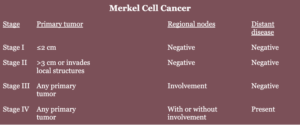
NCCN Staging
Treatment
- Wide local excision
- Margin: 1-2 cm
- Excise down to fascia or periosteum (level III evidence)
- No guidelines establish margins
- Sentinel lymph node biopsy (SLNB)
- Doesn’t reduce local recurrence rate
- Formal lymphoscintigraphy recommended for torso and head/neck Merkel cell cancer as drainage pattern is less predictable
- Negative SLNB → doesn’t preclude regional recurrence
- Positive SLNB → completion lymphadenectomy or radiation therapy
- Adjuvant radiation therapy
- Surveillance → skin exam and comprehensive nodal evaluation
- 3-6 months for 3 years
- 6-12 months for 3 years
- MCPyV should be evaluated every 3 months if patient has detectable level at the time of diagnosis. Consider imaging if titer increases >30% from prior lab value.
Resources
- STAMP Trial (2025)
- ADAM Trial (2026)