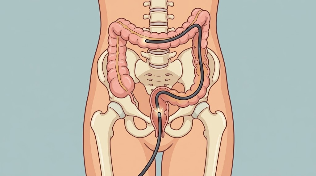
The flexible fiberoptic colonoscopy as used today was invented by William I. Wolff and Hiromi Shinya in 1969 in New York City. Prior to this, only a portion of the colon was routinely examined via rigid sigmoidoscopies. Today, colonoscopies are recommended as a routine method of screening for colon cancers starting at age 45 (a change from age 50 made in May 2018).
Indications
- Colorectal Cancer Screening Guidelines – United States Preventive Services Taskforce
- Colorectal Cancer Screening Guidelines – American Society of Colon and Rectal Surgeons
- Screening Tests to Detect Colorectal Cancer and Polyps – National Cancer Institute
- Screening for Colorectal Cancer – United States Centers for Disease Control and Prevention
Technique
- Preoperative considerations
- Hold anticoagulants 5-7 days before procedure
- Mechanical bowel prep completed by patient the day before procedure
- Surgeon holds endoscope in left hand, manipulating the deflection knob, suction, air, water with fingers while using the right hand to advance the length of the endoscope
- Lesion – when performing sampling, position the lesion at 5-6:00 to help with easier dissection
- Procedure
- Colonoscope is positioned in rectal vault and advanced when lumen is visualized. If lumen isn’t visualized, the colonoscope should be pulled back and insufflation should be performed with reassessment of the position of the scope.
- Looping of the colonoscope can occur and should be avoided. A straight colonoscope can reach the splenic flexure in 50-55 cm and the cecum in 100 cm, normally.
- Colonoscope can be advanced until the cecum is reached. During withdrawal, the instrument tip should be manipulated with small movements to allow for full visualization of the mucosa.
- Identifying areas of the colon
- Hepatic flexure → bluish cast due to liver
- Transverse colon → triangular folds
- Descending colon → long and straight, few haustrations
- Sigmoid colon → frequent turns
- Rectum → highly vascularized lumen
- Therapeutic interventions during endoscopy
- Snares (most commonly used)
- Endoscopic mucosal resection (EMR)
- Preferred for large, nonpedunculated lesions
- Goal to remove en bloc with 2-3 mm normal mucosa
- Local recurrence rate: 20% (piecemeal), 3% (en bloc)
- Endoscope submucosal dissection (ESD)
- Indications
- T1 carcinomas
- Technique
- Make a circumferential incision
- After incising half of the circumference, submucosal dissection is carried out
- Local recurrence rate: 1% (en bloc)
- Indications
Follow up
- Multiple factors go into follow up
- Recommendations for Follow-Up After Colonoscopy and Polypectomy (U.S. Multi-Society Task Force on Colorectal Cancer – March 2020)
Other Screening
- No test is perfect, and these are no exception. They are NOT diagnostic. They should be utilized in select patients. It is important to note if these tests come back positive, colonoscopy is recommended.
- Fecal immunochemical test (FIT)
- Guaiac-Based fecal occult blood test
- Cologuard
- CT colonography
- Flexible sigmoidoscopy
- mSEPT9
- Blood-based screening test for colorectal cancer
- Average risk adults ≥50 years old that declined screening colonoscopy or FIT
Complications
- Bleeding (4%)
- Perforation (0.4-2%)
- Postpolypectomy syndrome
- Surgical site infection
- Postoperative ileus