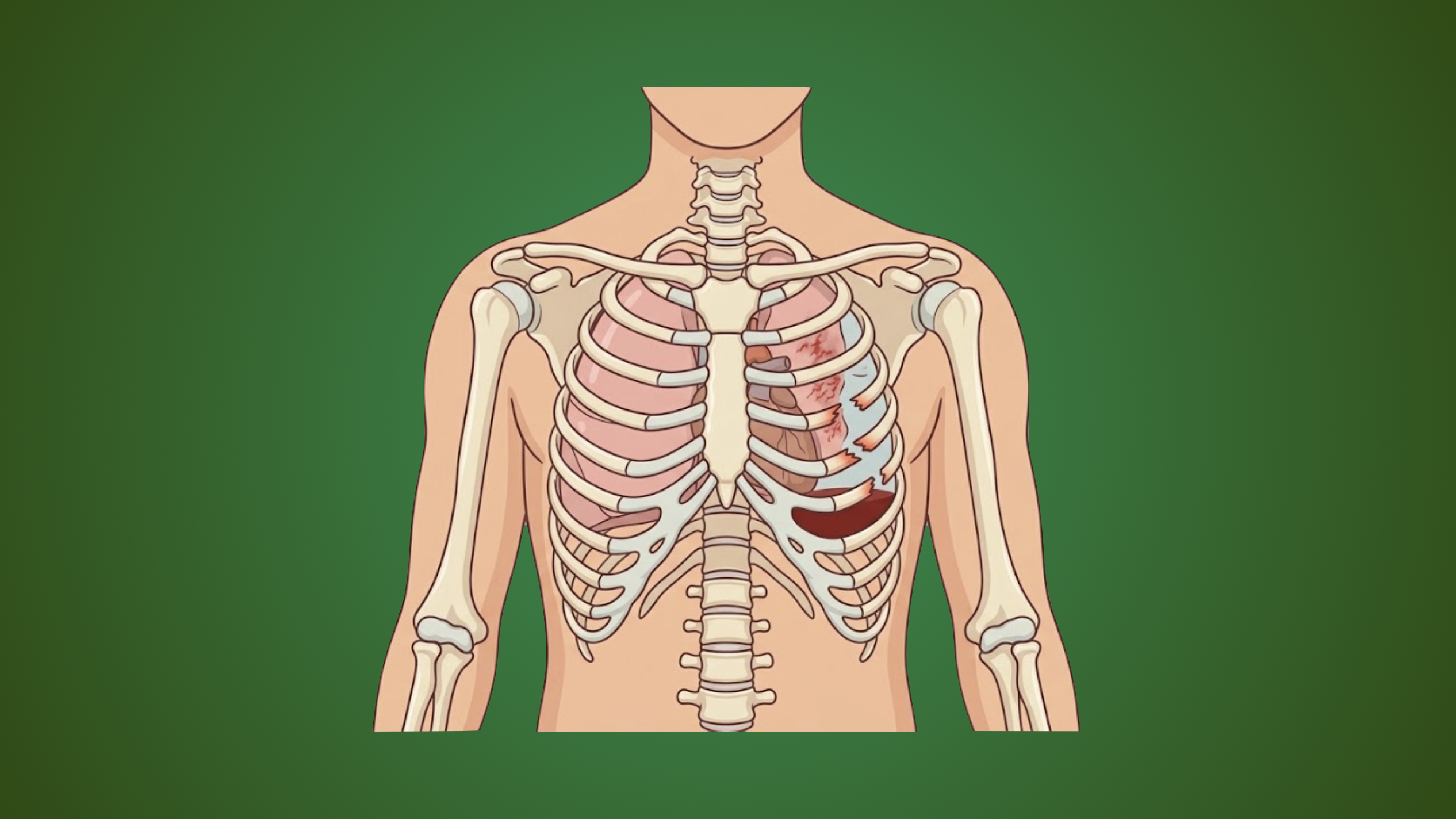
Thoracic trauma includes any damage to the chest, lungs, heart, great vessels, or the esophagus. It can be the result of blunt or penetrating trauma and accounts for 20-25% of all trauma-related deaths.
General Workup
- NEXUS indications for CXR
- Age >60 years
- Rapid deceleration mechanism
- Fall >20 feet
- Motor vehicle crash >40 mph
- Chest pain
- Intoxication
- Abnormal alertness or mental status
- Distracting injury
- Chest wall tenderness
- NEXUS indications for chest CT
- Abnormal CXR
- Distracting injury
- Chest wall tenderness
- Sternum tenderness
- Thoracic spine tenderness
- Scapula tenderness
- Rapid deceleration injury
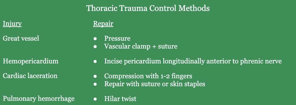
Thoracotomy
- Goals
- Control bleeding
- Relieve cardiac tamponade
- Cardiac compression
- Cross-clamp aorta → improve perfusion to brain/heart
- Indications
- Isolated penetrating chest wounds
- Blood loss >1500 mL after chest tube placement or blood loss >200 mL per hour for 4 hours
- Exposure
- Median sternotomy
- Heart
- SVC
- Pulmonary vessels
- Pulmonary hilum
- Left posteriolateral
- Posterior heart
- Thoracic structures
- Thoracic aorta
- Great vessels
- Left lung
- Distal esophagus
- Distal left mainstem bronchus
- Right thoracotomy
- Right lung
- Trachea
- Proximal mainstem bronchi
- Upper and midesophageal injuries
- Median sternotomy
Rib Fractures
- Presentation
- Most commonly fractured ribs: 4-10
- Tenderness to palpation
- Flail chest → segmental fractures of ≥3 ribs
- Paradoxical movement of chest with respirations
- Chest moves inward with inspiration and outward with exhalation
- Associated with other injuries due to blunt mechanism
- Scoring Systems
- RibScore → ≥4 in total may benefit from surgical stabilization
- ≥6 rib fractures
- ≥3 bicortical displaced fractures
- ≥1 fracture in each anatomic area
- Flail chest
- Bilateral fractures
- First rib fracture
- Rib Fracture Score
- Chest Wall Injury Score
- Clinical Rib Score
- RibScore → ≥4 in total may benefit from surgical stabilization
- Treatment
- Admission criteria for rib fractures
- Admission if ≥2 of the following present after PO pain medication
- Respiratory rate >18
- Incentive spirometry <75% predicted
- Numeric pain score ≥6
- Poor cough
- Age ≥65
- ≥3 fractures
- If admission criteria met, ICU admission if ≥1 of the following are present
- Age ≥65
- ≥6 fractures
- Incentive spirometry <60% predicted
- Flail chest
- >4L oxygen required to maintain SpO2 >90%
- Associated injury requiring ICU admission
- Admission if ≥2 of the following present after PO pain medication
- Multimodal pain regimen
- Oral
- Paravertebral nerve block
- Thoracic epidural anesthesia (gold standard for flail chest)
- Surgery
- Controversial
- Indications
- Crushed chest with chest wall deformity
- Flail chest
- Rib fracture nonunion with chest wall deformity
- Open rib fracture
- Intractable pain
- Persistent paradoxical respiratory movement
- If completed, should take place within 72 hours of injury
- Admission criteria for rib fractures
- Complications
- Pneumonia (most common)
- Atelectasis
Sternal Fractures
- Etiology: unrestrained driver striking steering wheel
- Presentation
- Severe chest pain
- Decreased respiratory efforts secondary to pain
- Associated injuries
- Myocardial contusion
- Rib fractures
- Thoracic spine fractures
- Workup
- Lateral CXR
- CT chest
- Treatment
- Medical management → pain control
- Surgery → create longitudinal incision overlying the sternum to expose fracture. Reduce overlying fractures by using a periosteal elevator and secure with a sternal plate
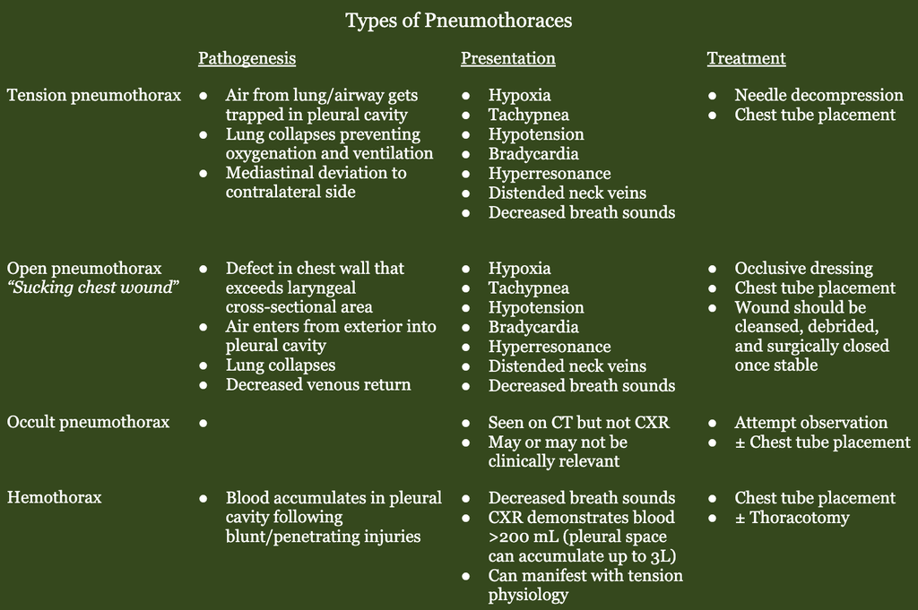
Pneumothorax
- Etiology
- Penetrating trauma
- Blunt trauma
- Presentation
- Decreased breath sounds
- Hyperresonance to percussion
- Decreased expansion of affected lung during inspiration
- Imaging
- CXR: collapsed lung
- CT: collapsed lung
- Treatment
- Surgical exploration indications
- Chest tube output >1500 mL initially
- Chest tube output >200 mL each hour for 4 hours
- Surgical exploration indications
Pulmonary Laceration
- Etiology
- Rib fracture
- Blunt or penetrating trauma
- Treatment
- Surgery
- Indication
- Initial hemothorax >1500 mL
- Hemothorax >200 mL/hour for 4 consecutive hours
- Persistent air leak for several days
- Approach
- Incision
- Posterolaterally through intercostal space 5-6
- Anterolateral through interspace space 4-5
- Repair
- Simple laceration → continuous absorbable suture
- Through and through penetration → pulmonary tractotomy with linear stapler
- Marginal injuries → wedge resection with linear stapler
- Hilar injuries → lobectomy or pneumonectomy
- Take down inferior pulmonary ligament with sharp dissection
- Grasp hilar structures between index and ring finger
- Clamp hilar structures
- Resect with TA stapler
- Incision
- Indication
- Surgery
Cardiac Injury
- Presentation
- Beck triad
- Hypotension
- Jugular vein distention
- Muffled heart sounds
- Hemorrhagic shock
- Beck triad
- Labs: Troponin
- Workup
- Ultrasound of pericardial space
- Subxiphoid pericardial window
- EKG
- Treatment → surgery
- Incision
- Median sternotomy
- Left anterolateral thoracotomy
- Injury and repair
- Atria
- Digital pressure or vascular clamp
- Close with continuous suture
- Ventricle
- Digital pressure
- Place Foley catheter and inflate balloon to 5-10 mL and outward traction
- Close with interrupted pledgeted horizontal mattress with monofilament nonabsorbable suture (i.e., 3-0 or 4-0 polypropylene)
- Can attempt closure with skin stapler
- Atria
- Evaluate after with TTE
- Incision
- Relevant Information
- “Box” boundaries —> cardiac injuries in 20% of patients
- Clavicles
- Midclavicular lines
- Costal margins
- Right ventricle is most commonly injuried structure of the heart
- “Box” boundaries —> cardiac injuries in 20% of patients
Myocardial Contusion
- Etiology
- Blunt thoracic injury
- Workup
- EKG
- Ongoing cardiac monitoring is indicated if EKG is abnormal
- TTE should be ordered if there is hemodynamic instability or myocardial contusion is suspected
Tracheobronchial Injury
- Etiology: Penetrating or blunt trauma
- Presentation
- Most occur within 2 cm of the carina
- Associated injuries
- Pneumothorax
- Pneumomediastinum
- Hemoptysis
- Subcutaneous emphysema
- Workup
- Flexible bronchoscopy (most accurate)
- CT chest
- Treatment
- Injuries <25% circumference
- Usually heal without stricture
- Don’t frequently require surgery
- Minimize inspiratory pressures when ventilated
- Surgery
- Approach
- Injury within 2 cm of carina → usually approached through right posterolateral thoracotomy incision
- Distal injury → left posterolateral thoracotomy incision
- Repair
- Primary repair with interrupted and continuous absorbable suture
- Tracheal (<4 cm) or mainstem bronchus (<2 cm) → resectional debridement with primary repair after mobilization, cover with vascularized pedicle (intercostal, diaphragm, or omentum
- Approach
- Injuries <25% circumference
Esophageal Injury
- Etiology
- Gunshot wound
- Stab wound
- Iatrogenic injury
- Presentation
- Usually identified during exploration for associated injury
- Dysphagia
- Hematemesis
- Crepitance
- Odynophagia
- Workup
- Endoscopy
- CT chest: mediastin/pleural air/fluid
- Treatment
- Surgery
- Approach
- Cervical esophagus → incision at anterior border of sternocleidomastoid
- Upper and middle thoracic esophagus → right posterolateral thoracotomy
- Distal esophagus → left 6th intercostal space posterolateral thoracotomy
- Tube thoracotomy is required
- Definitive treatment should be performed as soon as possible
- Repair
- Diagnosis within 24 hours → repair primarily with interrupted 3-0 or 4-0 sutures
- Delayed diagnosis → simple repair with reinforcement with intercostal muscle, pleural or diaphragmatic flaps, exclusion, diversion via a spit fistula, intraluminal T-tube drainage, endoscopic stenting, resection + drainage with NG tube or tube thoracotomy → contrast esophagram 5-7 days post repair prior to resuming PO intake
- Approach
- Endoscopic stenting gaining popularity slowly
- Surgery
Retained Hemothorax and Empyema
- See this Surgery Student post for more details
- Treatment
- VATS is most efficacious
- Most successful when completed within 3 days of injury as the clots becomes more adhered to the lung and pleura as days progress. It is largely unsuccessful when completed after 7 days.
- Place in lateral decubitus position
- Three 1-2 cm operative sites
- Posterolateral thoracotomy through 5th intercostal space if VATS is unsuccessful
- VATS is most efficacious
Diaphragm Injuries
- Etiology
- Penetrating trauma
- Risk factors: penetring injury between level of nipple and costal margin
- Epidemiology
- Male predominance
- Presentation
- Pain, dyspnea, respiratory failure
- Often enlarge with time
- Concomitant injuries to stomach, colon, liver are common
- Bowel and other organs can herniate through these defects, especially when large (>5 cm) and become incarcerated or strangulated
- Diagnosis
- Direct operative exam is gold standard – laparoscopy can be beneficial
- Imaging has a high false negative rate, especially for small (~1 cm) diaphragmatic defects
- Treatment
- Surgery
- Laparoscopic primary repair if hemodynamically stable
- 0 or 1-0 monofilament suture, running or interrupted
- Need for mesh for complex repair is quite rare (only use if clean)
- Surgery
- Relevant Information
- Blood supply to diaphragm
- Phrenic arteries
- Superior epigastric arteries
- Intercostal arteries
- Venous outflow: phrenic veins → IVC
- Blood supply to diaphragm