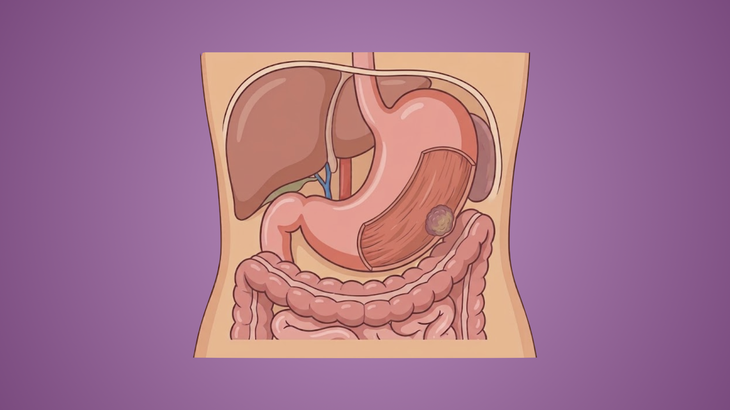
Gastrointestinal stromal tumor (GIST) is a rare cancer that develops in the wall of the digestive system called the interstitial cells of Cajal, which are the pacemaker cells that move food through the body. In 1983, Mazur and Clark introduced the term GIST after a gastric spindle cell tumor lacked smooth muscle differentiation. In 1998, it was discovered by Hirota and others that the GISTs originate from the interstitial cells of Cajal.
Etiology
- Sporadic (95%)
- Germline mutations
- Carney-Stratakis syndrome
- Carney triad
Epidemiology
- Age 65-70 years
- Found equally between males and females
Pathogenesis
- Originate from interstitial cells of Cajal
- Gain of function mutation at tyrosine kinase receptor (c-KIT)
- Histologic characteristics
- Spindle cell (70%)
- Epithelioid (20%)
- Mixed type (10%)
Presentation
- Median age: 60 years
- Median size: 5-7 cm
- Location
- Stomach: >50%
- Small bowel: 25-35%
- Majority are ileum and jejunum
- Duodenum: 5%
- Colon: 10%
- Rectum (5%)
- Esophagus (2%)
- Symptomatic (80%)
- GI bleeding
- Abdominal pain
- Asymptomatic (20%) → incidental finding
Workup
- Triple-phase contrast enhanced CT (CECT) A/P with PO and IV contrast
- Imaging of choice
- Brightly enhancing solid mass with smooth margins ± central necrosis and hemorrhage
- MRI → comparable to CECT
- Chest imaging
- Recommended for staging in all GISTs > cm
- Modalities
- CXR
- CECT chest
- EGD with EUS
- Preferred diagnostic method for foregut GISTs, especially submucosal nodules <2 cm. Biopsies should be taken endoscopically and not percutaneously due to risk of peritoneal dissemination.
- EGD: submucosal bulges with smooth margins and normal overlying mucosa +/- central ulceration
- EUS:
- Well-defined homogenous hypoechoic lesions originating from muscularis propria
- High-risk features
- Irregular borders
- Cystic spaces
- Echogenic foci
- Heterogeneity
- Ulcerations
- Immunohistochemistry + molecular characterization
- Gold standard for diagnosis
- KIT (CD117) and DOG1 → expressed in majority of cases
- CD34
- PDGFRA
- Usually diagnosed based on imaging and endoscopic findings
Staging
- AJCC
- Staging for gastric and omental GISTs is separate from the other GIST locations
- Distant metastasis is most common in liver and peritoneum
Treatment
- GISTs <2 cm and asymptomatic → possible observation
- Low risk of progression and metastasis
- Serial imaging with periodic endoscopic or other imaging
- GISTs >2 cm or symptomatic → surgery
- Standard of care; only modality that is potentially curative
- Goal for gross margin-free tumor (R0) with intact pseudocapsule. If there is a positive margin (R1), re-excision isn’t recommended. No lymph node dissection is needed.
- Approach
- Gastric GIST
- Greater curvature → stapled wedge resection
- Posterior wall → complete stomach mobilization followed by stapled or nonstapled resection
- Pylorus or GE junction →
- Resection over endoscope/bougie
- Stomach mobilization followed by transverse closure
- Distal or proximal gastrectomy, or subtotal gastrectomy can be performed if partial resection might compromise the function postoperatively
- Duodenal GIST
- Small tumors in first-second portion of duodenum → partial duodenal resection with primary closure
- Larger tumors → Roux-en-Y duodenojejunostomy
- Consider preoperative Imatinib if pancreaticoduodenectomy is required to obtain margin-free resection
- Tumors in third-fourth portion of duodenum → segmental duodenal resection with side-to-end or end-to-end duodenojejunostomy
- Jejunal and ileal GIST
- Localized, potentially resectable → en-bloc margin-free segmental bowel resection with primary anastomosis
- Rectal GIST
- Small GIST in distal rectum → transanal full-thickness excision with primary closure
- Mid-distal rectum →
- Transanal endoscopic microsurgery (TEMS)
- Transanal minimally invasive surgery (TAMIS)
- Upper rectum → anterior resection
- Larger tumors or tumors with difficulty with sphincter salvage → neoadjuvant therapy
- Gastric GIST
- Unresectable disease → preoperative tyrosine kinase inhibitors (TKIs) with repeat CT in 6-8 weeks followed by surgical resection and completion of target therapy
- Preoperative TKI indications
- Locally advanced tumors
- GISTS in duodenum, distal rectum, esophagus
- Evaluate for response to TKI with imaging (CT or MRI every 8-12 weeks)
- Response criteria
- RECIST
- Choi
- WHO
- Preoperative TKI indications
- Metastatic disease → medical therapy with TKIs (e.g., Imatinib)
- Postoperative care
- Surveillance
- Clinical evaluation every 3 months
- Cross-sectional imaging every 3-6 months for the first 5 years and annually thereafter
- Adjuvant therapy
- Surveillance
Relevant Information
- Most common mesenchymal tumor arising from GI tract → 1-2% of primary GI neoplasms
- Location of GIST
- Stomach (most common – 40-60%)
- Small intestine (20-30%)
- Colorectal (5-15%)
- Esophagus (2-5%)
- Less common: mesentery, omentum, pancreas, GU tract
- Risk stratification
- Based on primary site, tumor size, and mitotic rate
- Prognostic models
- Armed Forces Institute of Pathology (AFIP)
- National Institute of Health (NIH)
- Predictors of poor prognosis
- Size >5 cm
- Mitosis >5/50 high-powered fields
- Non-gastric site
- Usually benign, but can be malignant. The most common site of metastasis is the liver.
- Memory devices → GI5T for poor prognosis
- Size >5 cm
- Mitoses >5/50 high-powered fields