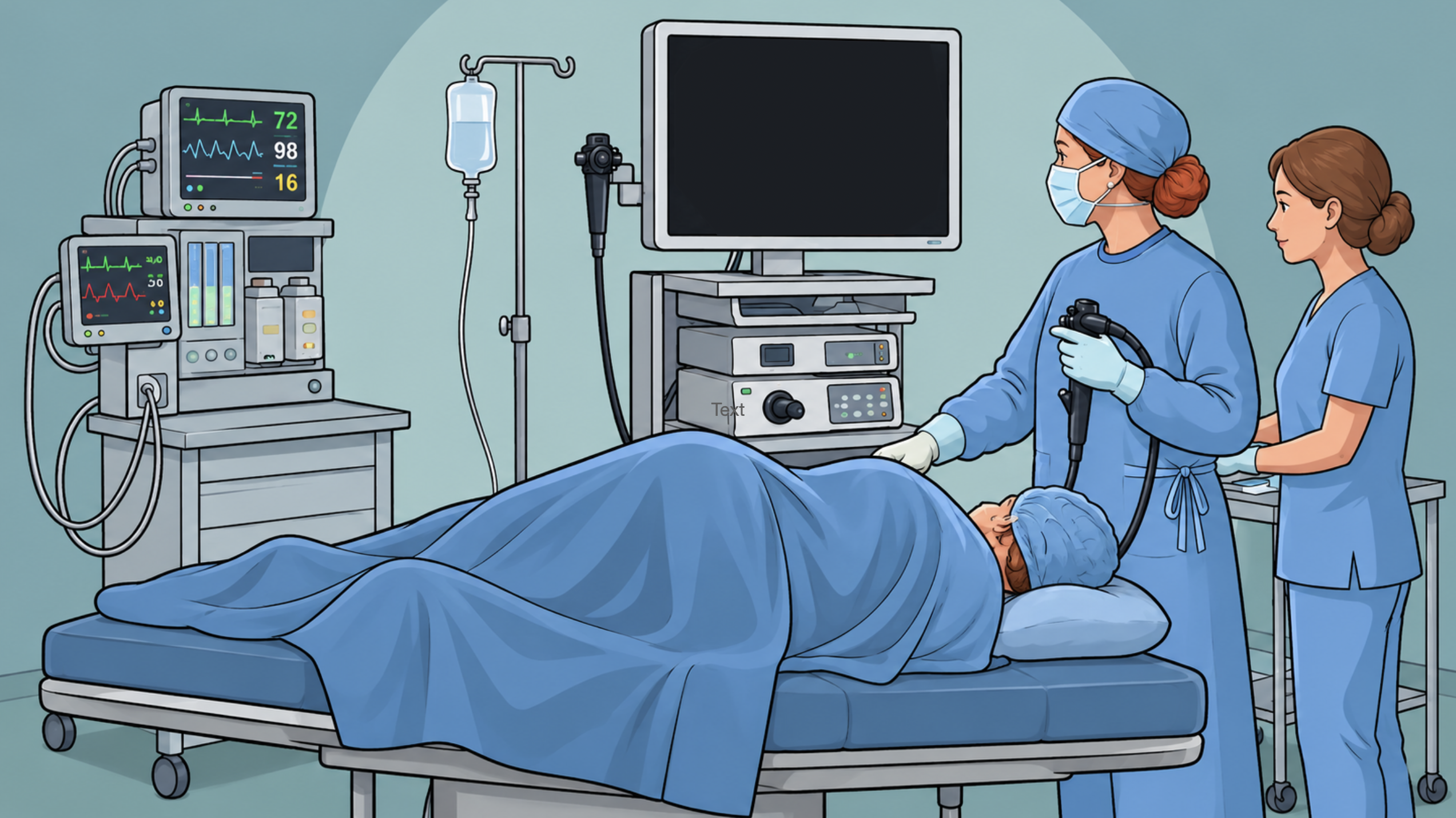
Upper endoscopy, or esophagogastroduodeoscopy, refers to a procedure in which a thin flexible tube with a camera and light are used to evaluate the upper digestive tract. In 1805, the first endoscope was created by Phlip Bozzini in Frankfurt. In 1853, the term “endoscopie” was coined when Antoine Jean Desormeaux created a gas-filled illuminated tube. Advances continued into the 1860s and 1900s until the modern endoscope was born.
Indications
- Dysphagia
- Odynophagia
- Pyrosis (heartburn)
- Suspected foreign body
- Abnormal barium radiograph
- Upper GI bleed
- Need for tube gastrostomy
- Persistent vomiting
- Unrelenting epigastric pain
- Gastric polyposis
- Surveillance for neoplasia with conditions that predispose to malignancy:
- Barrett esophagus
- Gastric ulcer
- Pernicious anemia
- Previous gastrectomy
Contraindications
- Absolute
- Severe comorbidities
- Inability to tolerate conscious sedation
- Relative
- Recent GI anastomoses
- Inflammation of the GI tract
Technique
- Stomach is empty
- No PO intake of solid food 8 hours pre-procedure
- No PO intake of clear liquids 2-4 hours pre-procedure
- Positioning: left side down
- Surgeon holds endoscope in left hand, manipulating the deflection knob, suction, air, water with fingers while using the right hand to advance the length of the endoscope
- Procedure
- Advance endoscope into esophagus under direct visualization – advance over the tongue until epiglottis and vocal cords are visualized, then angle the tip of the endoscope posteriorly between arytenoids with slight pressure to advance into esophageal introitus.
- Insufflate air after entering into the esophageal lumen and inspect mucus, noting peristalsis during exam. The stomach should be entered without delay initially to ensure there isn’t any retained food as this could result in aspiration and complications.
- Perform biopsy for any interruptions in normal mucosa or where there are areas of inflammation or suspected neoplasia.
- Insufflate the stomach with air until it is completely distended. Reduce and inspect mucosa in any areas where redundancy is noted. Orient the endoscope as below:
- 3:00 → posterior wall of stomach
- 6:00 → greater curvature of stomach
- 9:00 → anterior wall of stomach
- 12:00 → lesser curvature of stomach
- Advance endoscope toward gastric antrum by following the lesser curvature. Incisura angularis will be observed at 12:00 position and will appear as a smooth curved arch dividing the gastric body from the antrum. Retroflexion of the scope with slight withdrawal is performed to view the gastric cardia and fundus. Once it is no longer retroflexed, the cardia, fundus, and lesser curvature are then fully inspected.
- Pyloric orifice is visualized and endoscope is advanced into duodenal bulb and small movements are carried out to see small areas where ulcers can be difficult to identify. The endoscope is then advanced into the descending duodenum, which requires maneuvering the endoscope not under direct vision – turning scope posteriorly and down. The endoscope is then withdrawn. Ampulla of Vater may be seen at 9-12:00 position in descending duodenum.
- Therapeutic interventions during endoscopy
- Monopolar
- Bipolar
- Endoscopic clips
- Hemostatic sprays
- Sclerotherapy
- Rubber band ligation
- Hydrostatic balloons
- Endoscopic mucosal resection (EMR)
- Can remove esophageal and gastric lesions, including Barrett esophagus, dysplasia, and early carcinoma
- Often injecting saline in order to elevate the lesion followed by resection with a snare
- Additional techniques: suck-and-cut, suck-and-ligate
- Endoscopic submucosal dissection (ESD)
- Can remove early esophageal neoplasms, localized gastric cancers, and sometimes small duodenal cancers
- Technique
- Inject liquid into submucosal plane
- Divide mucosa and enter submucosal plane
- Complete resection with either needle cautery, blunt endoscope cap dissection, or a combination of the two
- Radiofrequency ablation (RFA)
- Stenting
- Per oral endoscopic myotomy (POEM)
- Performed for achalasia
- Enter submucosal space by creating submucosal bleb. Incise mucosa. Dissect submucosa by using a cap fixed to the tip of the endoscope. Then divide inner circular muscle fibers with an electrosurgical instrument in order to extend myotomy a few centimeters onto the stomach. Mucosal defect is closed with clips or endoscopic suturing